Download
1 / 52
990 likes | 2.08k Views
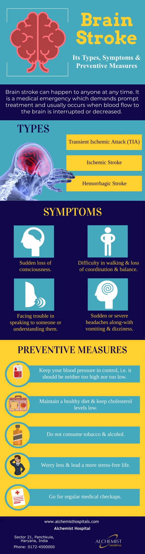
Types of Stroke. Objectives To review the two common types of stroke To review the stroke mechanism for the two common types of stroke To review the etiology of the two types of stroke To describe common patient presentations of stroke mimics. Ischemic (80%). Hemorrhagic (20%).

E N D
Types of Stroke • Objectives • To review the two common types of stroke • To review the stroke mechanism for the two common types of stroke • To review the etiology of the two types of stroke • To describe common patient presentations of stroke mimics
Ischemic (80%) Hemorrhagic(20%)
CT: Intracerebral Hemorrhage Intracerebral hemorrhage www.heartandstroke.ca/profed
Ischemic Stroke: Hyperdense MCA Sign Hyperdense MCA sign www.heartandstroke.ca/profed
Ischemic Stroke: Early CT Signs • Hyperdense middle cerebral artery sign • Subtle decreased attenuation of grey matter • Loss of grey-white differentiation • Loss of cortical ribbon • “Disappearing” basal ganglia • Early mass effect • Sulcal effacement • Shift www.heartandstroke.ca/profed
Ischemic Stroke: Etiology • Large Vessel Disease • Cardioembolic • Atherosclerosis • Small Vessel Disease • Lacunar Infarction • Cryptogenic
Secondary Vascular Malformations Aneurysms Tumors Hemorrhagic transformation of cerebral infarction Venous infarction with hemorrhage secondary to cerebral venous thrombosis Moya Moya disease Primary Chronic hypertension Cerebral amyloid angiopathy Anticoagulant/fibrinolytic use Antiplatelet use Drug use Other bleeding diathesis Intracerebral Hemorrhage: Etiology
Stroke Mimics • The following four conditions represent 62% of stroke mimics • Postictal deficit (unrecognized seizure) • Systemic infection • Tumour/abscess • Toxic-metabolic disturbance • Other mimics • Bell’s palsy • Peripheral nerve palsies • Old stroke • Confusion • Head trauma
Acute Stroke Management Resource Anatomy and Physiology Review
Objectives • Review the major blood vessels of the cerebral circulation • Anterior Cerebral Artery • Middle Cerebral Artery • Posterior Cerebral Artery • Review the key functional areas of the brain • List the common patient presentations related to carotid, vertebrobasilar and lacunar syndromes
Cerebrum Corpus Callosum • Largest portion • Two hemispheres • Joined by the corpus callosum • Dominance www.disenchanted.com/images/dictionary/corpus_callosum.gif
Right Hemisphere Spatial-perceptual deficits Left sided weakness/sensory loss Neglect of the affected side Distractible Impulsive behavior Poor judgment Loss of flow of speech Defects in left visual field-homonymous hemianopsia Left Hemisphere Expressive aphasia Receptive aphasia Global aphasia Right sided weakness/sensory loss Intellectual impairment- alexia, agraphia, acalulia Slow and cautious behavior Defects in right visual field-homonymous hemianopsia Left and Right Hemisphere
Cerebral Cortex • Divided into 4 lobes • Frontal • Parietal • Temporal • Occipital www.tbirecoverycenter.org/treatment.htm
Blood Supply to the Brain • Arterial supply from carotid and vertebral arteries which begin extracranially • Internal carotid arteries supply anterior 2/3 of hemispheres • Vertebral and basilar arteries supply posterior and medial regions of hemispheres, brainstem, diencephalon, cerebellum and cervical spinal cord www.heartandstroke.ca/profed
Circulation Review • Circle of Willis • Anterior Cerebral Artery (ACA) • Anterior Communicating Artery • Middle Cerebral Artery (MCA) • Posterior Communicating Artery • Posterior Cerebral Artery (PCA) Anterior Circulation Posterior Circulation
Anterior Cerebral Artery Anterior Cerebral Artery • Arises from internal carotid • Supplies anterior portion of basal ganglia, corpus callosum, medial and superior portions of frontal lobe and anterior parietal lobe • Key Functional Areas: • Primary motor cortex for leg and foot areas, urinary bladder • Motor planning in medial frontal lobe • Middle and anterior corpus callosum- communication between hemispheres www.cnsforum.com
Middle Cerebral Artery Middle Cerebral Artery • Arises from the internal carotid • Passes laterally under frontal lobe and between the temporal and frontal lobes • M1 segment- lentriculostriate arteries supply basal ganglia and most of internal capsule • Superior MCA branch- supplies lateral and inferior frontal lobe and anterior parts of parietal lobe • Inferior MCA branch-supplies lateral temporal lobe, posterior parietal and lateral occipital lobe www.cnsforum.com
Middle Cerebral Artery • Key Functional Areas • Primary motor cortex for face, arm and leg • Brocas language area (Superior MCA) • Wernickes language area (Inferior MCA) • Primary somatosensory cortex for face, arm, leg • Parts of lateral frontal and parietal lobes used in 3D visual-spatial perceptions of own body, outside world and ability to interpret and/or express emotions
Posterior Cerebral Artery Posterior Cerebral Artery • Blood supply for midbrain, hypothalamus and thalamus, posterior medial parietal lobe, corpus callosum, inferior and medial temporal lobe and inferior occipital lobe • Key Functional Areas: • Primary visual cortex • 3rd nerve in midbrain • Sensory control-temperature, pain, sleep, ADH • Communication between hemispheres www.cnsforum.com
www.strokecenter.org Posterior Cerebral Artery
Vertebrobasilar Circulation • Arise from the subclavian arteries • Run alongside the medulla • Blood supply for brainstem and cerebellum • Key Functional Areas: • Spinal cord tracts-pyramidal and spinothalamic • Cranial nerves 3-12 www.ib.amwaw.edu.pl/anatomy/atlas/image_12e.htm
Vertebrobasilar Circulation 1- Posterior Cerebral 2- Superior Cerebellar 3- Pontine Branches of Basilar 4- Anterior Inferior Cerebellar 5- Internal Auditory 6- Vertebral 7- Posterior Inferior Cerebellar 8- Anterior Spinal 9- Basilar www.ib.amwaw.edu.pl/anatomy/atlas/image_12e.htm
Cerebellum • Blood supply-own arteries from vertebrobasilar • Superior cerebellar • Anterior Inferior • Posterior Inferior • Major Functions • Control of fine motor movement • Coordinates muscle groups • Maintains balance, equilibrium www.daviddarling.info/images/cerebellum.jpg
www.answers.com Cerebellar Blood Supply
Brain Stem • Blood supply: PCA & Vertebrobasilar • Major divisions: midbrain, pons, medulla • Houses Cranial Nerves 3-12 • Serves as a pathway • Reticular Activating System
http://images.encarta.msn.com/xrefmedia/aencmed/targets/illus/ilt/T012872A.gifhttp://images.encarta.msn.com/xrefmedia/aencmed/targets/illus/ilt/T012872A.gif Cranial Nerves
www.colorado.edu/Kines/Class/IPHY3730/image/figure5-29.jpg Reticular Activating System
Collateral Circulation • Not all vessels have capability – lenticulostriate • Common sites: • External and internal carotid via opthalamic artery • Intracranial vessels of the Circle of Willis • Small cortical branches of ACA, MCA,PCA and cerebellar arteries
Collateral Circulation • Effectiveness depends on vessel size • Effectiveness depends upon speed of occlusion • Atherosclerosis • Circle of Willis: vessels are often narrow and cannot adapt for sudden onset of blockage
Collateral Circulation www.clevelandclinic.org/heartcenter/images/guide/disease/cad/artery7.jpg
Stroke Syndromes and Patient Presentations Acute Stroke Management Resource
Ischemic Stroke: Carotid Syndromes • Sensory/motor deficit • Aphasia • Cortical sensory loss • Apraxia, neglect • Retinal ischemia • Visual field deficit www.valleyhealth.com/images/image_popup/bn7_functionalbrain.jpg
Ischemic Stroke: Vertebrobasilar Syndrome • Diplopia • Vertigo • Coma at onset • Crossed sensory loss • Bilateral motor signs • Isolated field defect • Pure motor and sensory deficit • Dysarthria • Dysphagia www.state.sc.us/ddsn/pubs/head/brain.gif
Ischemic Stroke: Lacunar Syndromes • Makes up 25% of all ischemic strokes • Presumed to be occlusion of single small perforating artery • Predominantly in the deep white matter, basal ganglia, pons • Blood vessel: lenticulostriate branches of the Anterior Cerebral and Middle Cerebral Arteries • 30% of patients are left dependant and some long term data suggests up to 25% have a second stroke within 5 years (Wardlaw, 2007)
www.clevelandclincimeded.com/diseasemanagement/neurology/stroke/images/figure3.jpgwww.clevelandclincimeded.com/diseasemanagement/neurology/stroke/images/figure3.jpg Ischemic Stroke: Lacunar Syndromes
Case Examples • Add patient case examples of: • Anterior circulation strokes • Posterior circulation strokes • Lacunar Infarcts
Ischemic Stroke: Left (dominant) Hemisphere Stroke • Aphasia • Right field defect • Left gaze preference • Right upper motor neuron facial weakness • Right hemiparesis • Right hemisensory loss www.heartandstroke.ca/profed
Ischemic Stroke: Right (non-dominant) Hemisphere Stroke • Left neglect, inattention • Left field defect • Right gaze preference • Left upper motor neuron facial weakness • Left hemiparesis • Left hemisensory loss, sensory extinction www.heartandstroke.ca/profed
Ischemic Stroke: Cerebellar Infarct • Headache, nausea/vomiting • Vertigo, imbalance • Normal tone, power, reflexes • Inability to sit or stand • Ataxia • Late signs • Decreasing level of consciousness • Diplopia, gaze palsy • Ipsilateral V,Vll impairment www.heartandstroke.ca/profed
Ischemic Stroke: Brainstem Stroke • Decreased LOC • Crossed findings • Ipsilateral lower motor neuron facial weakness or sensory loss • Contralateral hemiparesis • Pupillary changes • Hiccoughs, vertigo • Bilateral motor findings • Diplopia, gaze palsies, intranuclear opthalmoplegia • Dysphagia • Dysarthria • Ataxia www.heartandstroke.ca/profed
Conclusions • Rapid assessment and triage key to optimal treatment • CT scan required to exclude hemorrhage • Knowledge of typical stroke symptoms key • Anatomical and etiological diagnosis necessary • Exclusion of stroke mimics vital
Resources • American Association of Neuroscience Nurses www.aann.org • American Stroke Association www.strokeassociation.org • Brain Attack Coalition www.stroke-site.org • Canadian Hypertension Education Program www.hypertension.ca/chep/en/default.asp • Canadian Stroke Strategy www.canadianstrokestrategy.ca • European Stroke Initiative www.eusi-stroke.com


![READ⚡[PDF]✔ Stroke of Luck](https://cdn7.slideserve.com/13156044/slide1-dt.jpg)