Download
1 / 36
360 likes | 634 Views
Rasha Buhumaid 200004343. Mr. A. A, 51 years old, Egyptian Chief complaint : vomiting blood and dark stool for the past few days. History of Present illness: he was doing well until 1 month ago Localized continuous dull Epigastric pain, severity 5/10. was partially relieved by zantac.

E N D
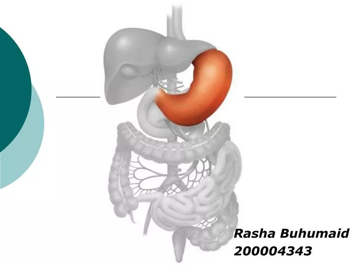
Rasha Buhumaid 200004343
Mr. A. A, 51 years old, Egyptian • Chief complaint: • vomiting blood and dark stool for the past few days.
History of Present illness: • he was doing well until 1 month ago • Localized continuous dull Epigastric pain, severity 5/10. was partially relieved by zantac. • Few days ago, vomit stained with blood (haematemesis) • Dark bloody stained stool (Melaena)
Past History: • Appendectomy 1983 • Was Treated for peptic ulcer 13 years ago. On Zantac PRN • Family History • Both parents are Diabetic and Hypertensive • Review of systems • Loss of appetite • Lost 6 kgs • Easily fatigued
What organs could be involved? • Esophagus • Stomach • Duodenum • Small intestine • Portal circulation
Physical examination • Heart Rate= 73 /min , • Blood Pressure= 110/80 mmHg • Afebrile • No pallor • No Jaundice
Abdomen examination • Symmetrical • Not distended • Scar of the appendectomy RIF • Abdomen was soft • Localized epigastric tenderness • No palpable mass / no organomegaly
What is your Differential Diagnosis? • Peptic ulcer (gastric, duedenal) • Portal hypertension • Gastric tumors • Duodenal tumors • Small intestinal tumors • Splenic vein thrombosis
What are the essential Blood Investigation • CBC • WBC and differential • CRP • Blood urea and electrolytes • Liver function test • Amaylase • Thyroid function test
Other Investigations • Upper GI endoscopy: • Mass with necrotic ulcer in the greater curvature of the stomach
Other investigation • Biopsy of the ulcer • Infiltrating adenocarcinoma • interstitial type • Poorly differentiated
Other Investigation • Serum Tumor Markers • CEA = 1.7 ug/l • CA 125 = 5.4 u/ml • CA 19.9 = 4.2 u/ml
Final Diagnosis Gastric Adenocarcinoma
Gastric adenocarcinoma • Epidemiology: • Twice as common in men • 2nd most frequent cancer but decreasing • Common in the 5th- 7th decades of life
Gastric adenocarcinoma • Risk Factors • Familial • Diet • salt, smoked, pickled food. • Lack of trace elements in the soil • Infection with H.Pylori • Precancerous conditions • Atrophic gastritis • Gastric polyps • Menetrier’s disease • Gastric ulcer • Previous gastric surgery • Pernicious anemia
Clinical presentation Primary: Obstructive symptoms (Dysphagia, vomiting) GI bleeding (Anemia, haematemisis, melaena) Perforation of lesion Abdominal mass Nausea Anorexia Weight loss Epigastric pain Asymptomatic Secondary: Abdominal distention Jaundice
Physical examination • Abnormal physical signs advanced disease • Supraclavicular lymph nodes enlargement • Jaundice • Ascites • Palpable epigastric/ pelvic mass
Essential Investigation • Blood test • CBC iron deficiency anemia • LFT liver metastasis • Serum markers (CEA, CA 19-9, CA 125) • Barium meal examination • Upper GI endoscopy and biopsy • Endoscopic ultrasound
Endoscopic Ultrasound EUS Image from the Stomach—Transducer (tr) is surrounded by a water-filled balloon (small arrows). Five layers of the gastric wall are visible. L = Liver. EUS image from the proximal stomach shows tumor (black arrows) with malignant lymph nodes (LN) also present. Liver (L) and diaphragm (white arrows) are also visible.
Other essential investigation • CT scan: pre-operative staging • Liver metastasis • Ascites • Pacreatic involvement
Other essential investigation • Laproscopy: • To enable direct inspection of the peritoneal cavity for metastasis (peritoneal seedings)
Pre-operative evaluation • Anemia should be corrected with blood transfusion • Electrolyte imbalance IV fluids • Nasogastric tube to empty the stomach • Single dose of cephalosporin 1 hr prior to surgery
Types of surgical procedures • Proximal third total gastroctomy with resection of distal 10cm of the esophagus • Large middle third tumor total gastroctomy • Distal third radical subtotal gastroctomy
Adjuvant therapy • Little evidence to support pre-post operative administration of chemotherapy. • Inadequacies of regimens
Early gastric cancer • Caners are confined to the mucosa and submucosa with strong evidence of staring of any lymph nodes involvement • Laser therapy • Endoscopic mucosal resection
Post operative consideration • NG tube & IV fluids are maintained until flatus has been passed and any paralytic ileus resolves. • Barium follow to exclude defects in reanastomosis technique.
Long term complication • Mictrocytic anemia (Fe deficiency) • Macrocytic anemia (Vitamin B12 deficiency) • Diarrhea • Dumping • Bile regurgitation