Download
1 / 52
530 likes | 1.28k Views
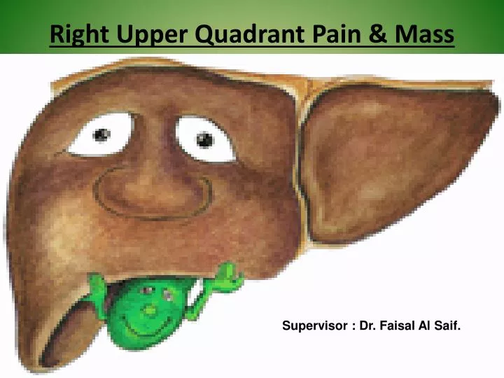
Right Upper Quadrant Pain & Mass. Supervisor : Dr. Faisal Al Saif. DDX. Gallstones. Acute cholecystitis. CA of gallbladder. Liver tumors. Liver abscess and cyst. Acute Cholecystitis:.

E N D
Right Upper Quadrant Pain & Mass Supervisor : Dr. Faisal Al Saif.
DDX • Gallstones. • Acute cholecystitis. • CA of gallbladder. • Liver tumors. • Liver abscess and cyst.
Acute Cholecystitis: - Severe RUQ Pain- Constant (Not Colicky)- Fever- +Ve Murphy’s Sign = Pt Will Stop Breathing In Deep Inspiration.- Increase WBC - Minor Elevation Of LFT- Bilirubin Is Normal. (Why)- no jaundice
Diagnosis US : Is Initial Study Of Choice In Most Patient With Biliary Disease Both Sensitivity And Specificity Are 95% The Wall Of GB Is ThickenedPericholecystic Fluid.(Appearing Black) Radiological Murphy's Sign (Put The US Prop Instead Of Ur Finger)HIDA scan : A Substance Injected Intravenously Will Be Taken By The Liver And Excreted As Bile Through The Common Bile Duct To Reach GB So, When The GB Is Absent In The Scan We Can Diagnose It As Acute Cholecystitis
Treatment - NPO - Analgesia- Antibiotics- Cholecystectomy Onset Of Symptoms> 72 Hours Immediate Surgery Onset Of Symptoms < 72 Hours Surgery Is Delayed For 4-6 Wks
GALLBLADDER TUMORS • Benign tumors • They are rare. • Papilloma. • Adenomyoma. • Fibroma. • Lipoma. • myoma. • myxoma, and carcinoid.
Malignant tumors • Carcinoma of the gallbladder. • 4% of all carcinomas of the gallbladder. It is the most common cancer of the biliary tract
CARCINOMA OF THE GALLBLADDER • 90% of the patients have cholelithiasis. • About 80% of the tumors are adenocarcinomas. • Metastases occur by lymphatic spread to the pancreatic, duodenal, and choledochal nodes and by direct extension to the liver.
RISK FACTORS • Gallstones large > small. • Porcelain gallbladder. • Cholecystenteric fistulas. • IBD • Mirizzi syndrome • Porcelain gallbladder : intramural calcification of the galbladder wall • Prophylactic cholecystectomy is recommended • Mirizzi syndrome refers to common hepatic duct obstruction caused by an extrinsic compression from an impacted stone in the cystic duct
CLINICAL PRESENTATION • Found incidentally at the time of cholecystectomy. • Right upper quadrant pain. • Jaundice and symptoms secondary to metastasis. • Associated with nausea and vomiting.
TREATMENT • If there is microscopic invasion of the gallbladder, cholecystectomy with wedge resection of the liver and regional lymphadenectomy may improve the survival. • Adjuvant chemotherapy is ineffective. • Radiation therapy is used to reduce tumor size and relieve jaundice. • 5-year survival rate is 0-10%.
HEPATOCELLULAR CARCINOMA • Most common primary tumor in the liver. • Incidence is 6 cases per 100,000. • Affects males more ,male :female ratio 2:1. • Average age is 50 years old. • Hepatocellular carcinoma occurs as a solitary mass or as multiple masses.
Local invasion, especially into the diaphragm, is common, as are distant metastases with the lung being most commonly involved.
RISK FACTORS • Chronic HBV and HCV. • Cirrhosis. • Metabolic disorders. • Schistosomiasis. • Environmental toxins. • Polychlorinated biphenyls. • Aflatoxins. • Thorotrast. • Alpha 1 anti tyrpsin deficiency.
CLINICAL PRESENTATION • Large lesions presents with a dull pain in RUQ malaise, fever,anorexia,weight loss and jaundice may be found. • On examination there will be hepatomegaly(88%),tender abdominal mass(50%),weight loss(85%)associated with cirrhosis(60%).
HOW TO DIAGNOSE • Alpha-fetoprotein increased in 75%. • Hepatic ultrasound • Not sensitive in lesions less than 2cm. • CT scan With contrast. • MRI
TREATMENT • Surgical treatment includes resection and transplantation. • Chemotherapy . Not given systematically. not given systematically
Combination therapy using chemoembolization and local ablation may be palliative for patients with unresectable lesions. • Ablative therapies include instillation of absolute ethanol into the lesion or insertion of a probe and delivery of radio frequency energy into the lesion.
CHOLANGIOCARCINOMA • a tumor that arises from the bile duct epithelium; it represents 5%–30% of all primary hepatic malignancies. • In most of patient it discovered incidentally.
CLINICAL PRESENTATION • Right upper quadrant pain, jaundice, hepatomegaly, and occasionally a palpable mass. Patients are usually 60–70 years of age. • Metastasis occurs initially to the regional lymph nodes or to the liver.
of intrahepatic tumors is resection when feasible. Overall survival is poor. Risk factors include parasitic infections (e.g., Clonorchis sinensis), primary sclerosing cholangitis, or Thorotrast exposure. Treatment
DIAGNOSIS • CT will show large hypovascular tumor with central necrosis.
METASTATIC TUMORS OF THE LIVER • liver is the second most common site of metastasis (exceeded only by regional lymph nodes) for all primary cancers of the abdominal viscera. • 60% of colorectal cancer. • 50% of cancers outside the abdomen. • 30% of all cancers ultimately spread to the liver,
which is the most common site of hematogenous spread. • Also from pancreas, breast, neuroendocrine and urogenital cancer.
HEMANGIOMA • Presentation : • Most common benign tumor 7-20 % . • Usually asymptomatic . • May Present with symptoms of compressing adjacent structure or stretching the liver capsule . • Right upper quadrant pain and mass and bruit. • There may be single or multiple masses . • Occur in female more than in male 3:1 .
INVESTIGATION • U/S . • CT with IV contrast . • Tagged RBC scan . • MRI . • Biopsy may cause hemorrhage . • * CT WITH IV CONTRASTDURING THE ARTERIAL PHASE ,THE TUMOR APPEARS AS A SHARPLY DEFINED MASS WITH SEQUENTIAL GLOBAL OPACIFICATION FROM OUTSIDE IN .
MANAGEMENT • Observation> 90% . • Surgical resection if there is symptoms or hemorrhage .
HEPATOCELLULAR ADENOMA • Presentation: • Female > male ( 9:1 ) between 30 – 35 years of age . • Associated with OCP and androgenic steroids . • Usually asymptomatic . • 25% have RUQ pain and mass . • Usually single but can be multiple in 30% of cases . • 30% present with spontaneous rupture and hemorrhage . • Risk of developing carcinoma .
INVESTIGATION • LFT : normal . • U/S . • MRI with gadolinium enhancement . • CT scan . • Biopsy : normal hepatocyte without bile duct . • * CONTRAST ENHANCED CTDEMONSTRATE MODERATE ENHANCEMENT DURING THE ARTERIAL PHASE SHOWS A NEARLY HOMOGENEOUS, WELL-ENCAPSULATED MASS
CT MRI
MANAGEMENTStop OCP, avoid pregnancy . • Observation : • Small , intrahepatic associated with OCP use . • Surgical resection : • Large >5cm , superficial , women anticipates pregnancy near .
FOCAL NODULAR HYPERPLASIA • Presentation : • Female > male approximately 40 years of age . • Associated with OCP . • Usually asymptomatic . • 10% have RUQ pain and mass . • Single or multiple lesion with nodular appearance
INVESTIGATION • U/S . • Contrast enhanced CT . • MRI with gadolinium enhancement . • Technetium 99m-labeled sulfur colloid scan .
Biopsy : The tumor contain hyperplastic hepatocyte with inflammatory cells and bile duct epithelium . * CONTRAST- ENHANCED CTAPPEARS AS HOMOGENOUS HYPER ATTENUATING IN ARTERIAL PHASE WITH CENTRAL SCAR AND RADIATING BANDS
MANAGMENT • stop OCP . • Observation . • Surgical resection or embolization .
Liver abscess and cysts • Bacterial • Parasitic • Fungal
Source • Direct spread from biliary tract infection . • Portal spread from GI infection . • Systemic infection . • Liver trauma . • 10-50% Cryptogenic .
PRESENTATION • RUQ pain and mass . • Tenderness over RUQ . • Systemic symptoms : • Fever , chills , sepsis , malnutrition , anemia .
INVESTIGATION • CBC : anemia , lukocytosis . • LFT: raised liver enzymes mainly alkaline phosphatase . • Indirect heamgglutination antibody test for parasite . • U/S . • CT .
MANAGEMENT OF BACTERIAL ABSCESS • IV antibiotics . • Pericutanous drainage with CT or U/S guidance . • Surgical operative drainage is indicated for multiple abscess or multiple pericutanous drainage have failed
HYDATID CYST • Result from infection with the parasite echinococcus granulosus . • Dogs are the definitive host shedding ova in the feces which infect intermediate host such as man , sheep , cattle . • Endemic infection .
May progressively enlarged and ruptured : • 50% within hepatic parenchyma form daughter cysts . • Bile duct : debris cause biliary obstruction . • Peritoneal cavity : urticaria , esinophilia , anaphylactic shock .