Download
1 / 37
670 likes | 2.05k Views
Assessment of Musculoskeletal System. By Dr. Hanan Said Ali. Learning Objectives. Explain how to assess musculoskeletal system. Musculoskeletal Anatomy and Physiology. Anatomy and physiology The musculoskeletal system comprises

E N D
Assessment ofMusculoskeletal System By Dr. Hanan Said Ali
Learning Objectives • Explain how to assess musculoskeletal system
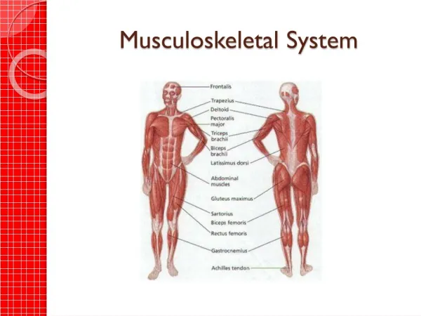
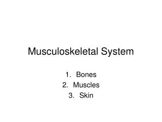
MusculoskeletalAnatomy and Physiology Anatomy and physiology The musculoskeletal system comprises bones, muscles and joints, and makes up most of the body’s mass. It performs a number of essential functions including: • Maintenance of body shape. • Support and protection of soft tissues structures such as the brain, heart and lungs.
Movement. • Breathing. • Storage of calcium and phosphate in bone. • The manufacture of red blood cells, white blood cells and platelets in the bone marrow (haematopoiesis)
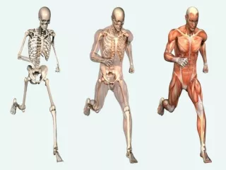
Bones • Bone is a rigid structure ideally suited for its supportive and protective function. Bones contain sites for muscle attachment, the mechanical basis for movement. Muscles • Muscle tissue is made up of contractile cells which have the ability to shorten in length or contract. It is this characteristic that is responsible for movement,maintenance of posture and heat production
Joints • A joint is the site at which two or more bones are united, providing the allows movement. • Fibrous joints unite bones by fibrous connective tissue and allow very little movement.
MusculoskeletalAssessment • Health History • Subjective Data -Chief Complaints – pain, altered sensation, limited motion • Family history, personal history, dietary history, socioeconomic status • Medications (steroids); Current health problems - obesity • Objective Data - Physical Exam • Objective Data - Diagnostic Tests
If there is alteration:Determine how alteration influences ability to perform activities of daily living (e.g., bathing, feeding, dressing, toileting, and ambulating)Social functions (e.g., household chores , work , recreation, sexual activities).
Physical Exam • Mental Status • General inspection Looks at the extremities for overall size , gross deformity , bony enlargement, ,and symmetry. • Head and neck: crepitus • Height, weight, nutritional status, skin, • spine – lordosis(is an increased lumbar curvature), scoliosis( Lateral spinal curvature), Kyphosis ( is an exaggeration of the posterior curvature of the thoracic spine).
Posture, gait, ROM ex., muscle strength and tone, size symmetry of muscle development .PalpationThe nurse applies gentle palpation to all bones, joint, and surrounding muscles to :Heat, tenderness, edema, or resistance to pressure.Normal* The client should feel no discomfort when palpation is applied.* Muscles should be firm.
Physical Exam (cont.) • A Tremor: is an involuntary trembling of a limb or body part. * Tremor may involve large groups of muscle fibers or small part. • An intention tremor: when an individual attempts a voluntary movement, such as holding a cup of coffee. • A resting tremor: is more apparent when the client is at rest and diminishes with activity. • Body posture is assessed for normal standing and sitting position.
Physical Exam (cont.) Bones are assessed for normal form. Joints are assessed for • tenderness • swelling , • thickening, • crepitation (the sound of bone grating on bone), • presence of nodules, • range of motion
Muscle Assessment • Inspection of the muscles: • For the size. Compare the muscles on one side of the body to the same muscle to the other side. For any discrepancies, measure the muscle with a tap. Normal finding Equal size on both sides of body Deviations from normal Atrophy (a decrease in size) or hypertrophy (an increase in size) asymmetry
Muscle Assessment (cont.) • Inspect the muscle and tendons for contractures ( shortening ). Normal finding No contractures Deviations from normal Malposition of body part ,e.g., foot drop (foot flexed downward. • Inspect the muscles for tremors , for example by having the client hold the arm out in front of the body Normal finding No tremors Deviations from normal Presence of tremors
Palpate the muscles for: • Palpate the muscles at rest to determine the muscle tonicity ( the normal condition of tension, or tone, of a muscle at rest). Normal finding Normally firm Deviations from normal A tonic (lacking tone).
Palpate muscle while the client is active and passive for : • flaccidity. • spasticity. • smoothness of movement. Normal finding Smooth coordinated movement Deviations from normal Flaccidity (weakness of laxness) or spasticity (sudden involuntary muscle contraction).
Muscle Assessment (cont.) Test muscle strength Compare the right side with left side. Sternocleidomastoid: • Client turns the head to one side against the resistance of your hand. • Repeat with the other side. Normal finding Equal strength on each side Deviations from normal 25% or less of normal strength. Applied for all muscles
Trapezius: Client shrugs the shoulders against the resistance of your hands . Deltoid: Client holds arm up and resists while you try to push it down. Biceps: Client fully extends each arm and tries to flex it while you attempt to hold arm in extension
Triceps:Client flexes each arm and then tries to extend it against your attempt to keep arm in flexion.Wrist and finger muscles: Client spreads the fingers and resists as you attempt to push the fingers together.Grip strengthClient grasp your index and middle fingers while you try to pull the fingers out.
Hip muscles:Client is supine, both legs extended ; client raises one leg at a time while you attempt to hold it down.Hip abduction.Client is supine, both legs extended. Place your hands on the lateral surface of each knee; client spreads the legs apart against your resistance.Hip adductionClient is in same position as for hip abduction. place your hands between the knee; client brings the legs together against your resistance
Bone AssessmentInspect : the skeleton for normal structure and deformities.Palpate : the bones to locate any areas of edema or tenderness.Normal findings: No deformities.No tenderness or swelling.Abnormal findings:Bones misaligned.Presence of tenderness or swelling(may indicate fracture, neoplasm, or osteoporosis
. Assessment of joints:Inspect : the joint for swelling.Palpate : each joint for tenderness, smoothness of movement ,swelling, crepitation, and presence of nodulesNormal findings:No swelling.No tenderness, swelling , crepitating or nodules.Joints move smoothly.Abnormal findings:One or more swollen joints.Presence of tenderness, swelling, crepitating , nodules.
Assess joint range of motion. Ask the client to move selected body parts. The amount of joint movement can be measured by a goniometer, adevicethat measure the angle of the joint in degrees. Goniometers are the measuring devicesGoniometer pin is placed over the joint and two arms are aligned with the limbs connected to the joint
Assess joint range of motion. (cont.)A proficiency anatomical landmark location aids in more accurate measurements. Best results are obtained if the joints are ranged slowly and smoothly.Two people should perform the test.One will range the joint and the other will hold and read the goniometer
Assess joint range of motion. (cont.)Normal FindingVaries to some degree in accordance with person’ genetic makeup and degree of physical activity.Deviation From NormalLimited range of motion in one or more joints
Diagnostic Evaluation • Imaging Procedures – CT, Bone Scan, MRI • Nuclear Studies - radioisotope bone density, • Endoscopic Studies –arthrocentesis, arthroscopy • Other Studies –biopsy, synovial fluid, Arthrogram, venogram, • Electromyography • Myelography* • Laboratory Studies