Download
1 / 66
660 likes | 915 Views
Cell Communication. Cell Signal, Transduction and Response. Signaling is Important.

E N D
Cell Communication Cell Signal, Transduction and Response
Signaling is Important • Just as we communicate with other humans in a number of different ways, cells communicate with other cells and with their external environment with a set of cell signal mechanisms and process signal information in order to make appropriate responses within the cellular environment.
Signaling is Complicated • Cellular communication is necessary to coordinate the myriad activities needed for any organism (unicellular or multicellular, prokaryote or eukaryote) to grow, develop and function. • Most organisms use the same kinds of cell signaling mechanisms affirming again the uniformity of DNA for life processes.
Signaling is Diverse • Communication of Self • Signals and receptor Proteins • Signaling Nearby and Signaling Far Far Away
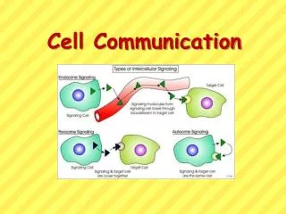
What are the Types of Signaling Methods? • Autocrine • Direct Contact • Paracrine • Synaptic • Endocrine
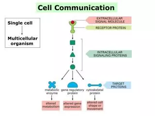
The Signal Transduction Pathway • Overview Animation • Reception: • A signal binds to a specific cellular protein called a receptor which is often on the surface of the cell. • Transduction: • The binding of the signal triggers a change in the receptor in some way (shape) which converts the signal into some sort of cellular response • Response: • The response can be almost any cellular activity such as activation of an enzyme or gene expression
Examples • Epinephrine stimulates vasodilation or vasoconstriction • Epinephrine also acts as a trigger for the cell to break down glycogen
Receptors • In General • Types: • Cytoplasmic • Gene Regulators • Enzyme Receptors • Plasma Membrane
Messengers • In General • Relay Proteins • Secondary Messengers • cAMP • Ca++ • Nitric Oxide
Signal Amplification • In General • Signal Efficiency • Signal Specificity and Response • Signal Regulation
Communication of Self • Cell recognition proteins are important to the immune system. • They play a critical role in communicating our identity to cells of our immune system and to our tissues. • Each cell has unique surface identity markers. Many of these are glycolipds, glycoproteins or a group of proteins called MHC (major histocompatibility complex) proteins. • The markers that form the human A, B and O blood groups are examples of identity markers. Most of the MHC proteins are immunoglobulins.
Signals and Receptor Proteins • Receptor proteins have attachment sites for our chemical messenger molecules or signal molecules. • Receptor proteins are specific; each "fits" a specific chemical messenger molecule. • The binding of the signal molecule to its protein receptor induces a conformational change in the receptor protein that ultimately leads to a response within the cell. • This sequence of cellular events is known as a signal transduction pathway. • Not surprisingly, the typical cellular response to a signal is gene regulation or enzyme activity. • Individual cells have different receptor proteins. This means that cells uniquely respond only to the signal molecules that are appropriate.
Signaling Nearby and Far Away • A chemical signal that communicates between two nearby cells is called a LOCAL REGULATOR • One type is called a paracrine signal • The other is called a synaptic signal • A chemical signal that communicates between cells some distance apart is called a HORMONE • A particular signal can act as both a local regulator or a hormone (ex Insulin)
Insulin • Insulin can interact with other pancreas cells to inhibit further insulin release or it can act on liver cells allowing the cells to start storing carbohydrates as glucagon.
Synaptic A specialized paracrine signaling occurs with cells of the nervous system. Neurotransmitters released at the axon end of one nerve cell traverse the space (called the synaptic cleft) to the target cells (receptor cells, nerve cells, or the neuromuscular junction).
Endocrine (Distance) Signaling Endocrine signaling involves chemicals produced in one cell or tissue that travel through the organism to their target cells and tissues. Many of these signal molecules are our regulatory hormones.
Receptors • A receptor can be located in the plasma membrane or within the cytoplasm of the cell. • A signal molecule binds to a specific site on its receptor. LIGAND is a term used to describe any (generally) small molecule that binds to a larger molecule. • Signal molecules are LIGANDS. • The signaling molecule has a shape that fits into a portion of its receptor protein, just as a key fits into a specific lock so a door can be opened. • Many signal molecules are polar and their receptor molecules are found within the plasma membrane. • Small nonpolar signal molecules, including many hormones and the gas, NO (nitric oxide), pass through the plasma membrane and have intracellular receptors. • Binding of signal molecules to their receptors is reversible, and critical, since a signal molecule that remained attached to its receptor would be constantly activating the receptor. • Although receptor molecules are specific to their signal molecules, inhibitors or antagonists can bind to receptors, blocking the receptor from functioning. • Campbell Animation • Agonists and Antagonists
Agonists and Antagonists • Not every ligand that binds to a receptor also activates the receptor. • Agonists are able to activate the receptor and result in a maximal biological response. • Most natural ligands are full agonists. • Partial agonists do not activate receptors thoroughly, causing responses which are partial compared to those of full agonists. • Antagonists bind to receptors but do not activate them. • This results in receptor blockage, inhibiting the binding of other agonists.Inverse agonists reduce the activity of receptors by inhibiting their constitutive activity.
Cytoplasmic Receptors • Cytoplasmic receptors function with small nonpolar signal molecules that can readily pass through the plasma membrane. • The receptors may be in the cytoplasm or in the nucleus. Intracellular receptors are often enzymes or gene transcription factors. • Gene Regulator CytoplasmicReceptors: Many of our steroid hormones function as signals for gene regulator receptors. • These receptors have specific DNA binding sites that are normally not accessible for transcription in the nucleus. • When activated by the signal molecule, the receptor complex is altered so it can function in the nucleus as a transcription factor to initiate gene activity.
Cytoplasmic Receptors • Some enzymes require signal molecules to become active. • Signal molecules function much the same way that co-factors and coenzymes work. • Each has a binding site on the enzyme that alters the conformation of the enzyme so that the target substrate can "fit". • Many of our digestive enzymes are produced in inactive forms and must be activated by signal molecules in the target location of the digestive system. • Nitric oxide (NO), which functions to relax smooth muscle tissue, is an enzyme signal molecule.
Plasma Membrane Receptors • A signal molecule that has a plasma membrane receptor will have a shape that fits into a portion of its receptor protein, which is an integral protein, in the plasma membrane. • The signal transduction is initiated in the membrane when the receptor protein reacts with its signal. • Sometimes the signal molecule promotes a conformational change in the receptor molecule that activates the receptor to interact with a specific cellular molecule, the responder, or an aggregation of receptor proteins, or activates a series (or relay) of molecular interactions leading to a cellular response. • In some cases, the ligand (signal molecule) promotes an aggregation of receptor proteins in the plasma membrane.
Types of Plasma Membrane Receptors • Ion Channel • Protein Kinase • G Proteins
Ion Channel • Ion channels are gated protein pores in the plasma membrane that open and close in response to signals. • The pores are highly specific and allow the flow of a specific molecule (typically Na+, Ca++ or K+). • Nerve transmission and muscle contraction rely on gated ion channels which, when open, rapidly cause a change in ion concentration as the ions flow through the pore. • This change in the polarity of the cytoplasm triggers a cell reaction or relay reactions.
Protein Kinase • Protein kinase receptors are in a group of proteins that have enzymatic activity. • A KINASE is a phosphorylating enzyme that catalyzes transferring phosphates from ATP to some specific protein. • Phosphorylatingsupplies energy.Tyrosinekinase membrane receptors are common in animal cells. • Serine and threoninekinases are also found in the cytoplasm of cells. • Protein kinase receptors catalyze phosphorylation of a region of the receptor protein when the signal molecule attaches to its surface. • Relay proteins (often protein kinases themselves) are then activated to elicit the appropriate cellular responses.Proteinphosphatase enzymes remove the phosphate molecules to de-activate the kinase relay molecules to turn off the pathway. • The phosphatase and kinase balance acts like an on-off switch for cellular pathways that rely on phosphorylations. • The tyrosine kinase receptor molecule consists of small alpha helix chains of tyrosine attached to the inactive enzyme "tail" on the cytoplasmic side of the membrane, and to signal binding sites on the extracellular side of the membrane.
Protein Kinase • When a signal molecule attaches to the binding site of its tryosinekinase receptor it triggers two tyrosine polypeptides to aggregate, forming a dimer. • The dimer conformation promotes the phosphorylation of the tyrosine molecules of the opposite polypeptide in the dimer. • Each polypeptide is catalyzing the phosphorylation of the tyrosines of the opposite dimer component. • The activated receptor is recognized by a number of relay proteins within the cell that undergo conformational changes when activated by the phosphorylatedtyrosines. • Multiple relay proteins can be activated at once so that a number of reactions can occur simultaneously within the cell. • The ability to elicit multiple responses is a characteristic of the protein-kinase receptors.
Examples • About 2% of human genes code for protein kinases. • Our cells may have hundreds of different protein kinases. • Insulin is a protein kinase signal molecule as are many growth factors. • The cyclin-CDK complex signals for cell division involve protein kinase receptors. • Some cancers may be caused by tyrosine-kinase receptors that aggregate (hence get phosphorylated) without their signal molecule, or by a defective phosphatase that may keep a relay continually phosphorylated (active).
G-Protein Coupled Receptors • There are a number of membrane receptor molecules that work with a special group of helper proteins called G-proteins (guanine-proteins). • The membrane receptor proteins that work with G-proteins have a common structure. • Each is comprised of 7 alpha-helices (a motif) within the membrane, an attachment site for the G-protein on the cytoplasmic side and an attachment site for the signal molecule on the extracellular side of the membrane.
How Do G-Proteins Work? • G-proteins are intermediates in cell signal pathways between receptor molecules and target molecules, which are often enzymes. • In their non-active form, G- proteins have guanine diphosphate (GDP) attached. • The active form of a G-protein has guanine triphosphate (GTP). ATP is used to phosphorylate GDP to form GTP. • G-proteins also have a binding site for the receptor protein in the plasma membrane and to an effector protein for the transduction path to effect a cellular response. • A ligand attaching to the receptor molecule triggers (by inducing a conformational change in) the receptor molecule to bind to its associated G-protein. • The receptor- G-protein complex triggers GTP to displace the inactive GDP on the G-protein. • The signal then dissociates from the receptor. • In general, a portion of the activated G-protein then migrates along the membrane and binds to a specific effector protein, typically an enzyme or an ion channel in the membrane. • The activated effector initiates a signal pathway in the cell resulting in a specific cell response. • The cellular response may be an activation or an inhibition.
GTP and G-Protein • GTP activation is short-term. • Once the G-protein activates its effector protein, GTP is hydrolyzed to GDP inactivating the G-protein and the G-protein subunits are re-associated to be ready to activate again. • The GTP hydrolysis back to GDP in the cell is catalyzed by the GTPase enzymatic activity of the G-protein. • (A G-protein serves as its own enzyme to catalyze the reaction of GTP to GDP.) This prevents chemical reactions from occurring in the absence of the appropriate signal molecule.
G-Protein Receptors • There are over 100 G-protein linked receptors. Although each is specific in function, they are closely related in structure. • Different subunits of the same G- protein may respond to the same signal in different tissues eliciting an activation in one tissue and an inhibition in a second, as in the response in different tissues to epinephrine, a stress (fight or flee) signal molecule. • G-proteins are important in: • genetic-gender reproduction • neurotransmitter function • sensory reception (vision, taste and smell) • embryonic development • hormone signaling
Signal Transduction pathways • In General • Relay Proteins • Secondary Messenger Molecules • cAMP • Ca++ • Nitric Oxide • Signal Amplification
Signal Transduction Pathways • Signal transduction would be “easy” if each signal molecule had a receptor that caused a direct cell response. • Such interactions between the signal and receptor in which the receptor causes the response are known as direct transduction. • However, the transduction process (translation of a signal) is more commonly an indirect transduction, involving cytoplasmic secondary messengers that mediate added steps in the transduction.
Signal Transduction Pathways • Both direct and indirect transduction, with a secondary messenger involved, can result in a cascade of steps, or transduction relays. • Pathways can provide more opportunities to coordinate and regulate cell activities and can also serve to amplify responses. • The proteins involved in these pathways are called relay proteins because they are "relaying" the information from the signal to the target response. • They also frequently serve to amplify the original signal to get a greater response.
Relay Proteins (Protein KinasePhosphorylation Cascade) • Many signal transduction pathways use a sequence of steps (or relay) to transmit the signal message within the cell. • The typical relay proteins are protein kinases (recall that a kinase is an enzyme that phosphorylates its substrate) that catalyze the phosphorylation of two amino acids, threonine and serine, on the next relay protein in the cascade, which in turn phosphorylates the next protein in the relay. • Each protein in the cascade, when phosphorylated, catalyzes the next. Ultimately one reaches the end of the pathway, or a branch in the pathway, for an appropriate cellular response. • Each cascade can amplify and communicate the needed signal until the final target is reached. • With different target proteins within the cascade, variable responses are also possible with phosphorylation cascades. • The protein kinase receptors are typically involved in direct transduction activating a phosphorylation relay (or cascade) because the receptor, once phosphorylated transfers its phosphate to the first protein in the relay.
Regulation • Protein-kinase cascades are common in growth and development activities and are often activated by growth factors. • Although we associate the protein kinase relay pathways with a response in the cell that promotes a reaction, it is also important to note that the pathway can work to de-activate rather than activate, diminishing cell activity. • That can be the appropriate response.Proteinkinase activity is regulated by feedback mechanisms. • A second set of proteins, the protein phosphatases, remove phosphates from protein kinases, stopping their activity. • Protein phosphatases are active when the signal molecule for a protein kinase is absent, which shuts down that particular signal transduction pathway.
Secondary Messenger Molecules • Membrane proteins are primary receptors for signal molecules, but many receptors require additional molecules in the cytoplasm in order to relay their message. • Small water-soluble molecules and ions can relay messages from the membrane proteins rapidly throughout the cytoplasm by diffusion. • These relay molecules, called secondary (or second) messengers, work with both G-protein receptors and protein kinase receptors.