Download
1 / 99
1.01k likes | 1.25k Views
The Skeleton: Bones & Joints. Chapter 7 Anatomy & Physiology I. Outline . BONES Main functions Bone structure Bone growth and repair Bone markings BONES OF THE AXIAL SKELETON Framework of the skull Framework of the trunk BONES OF THE APPENDICULAR SKELETON Upper division

E N D
The Skeleton: Bones & Joints Chapter 7 Anatomy & Physiology I
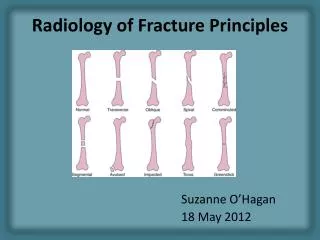
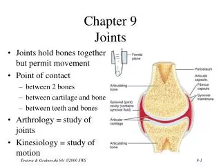
Outline • BONES • Main functions • Bone structure • Bone growth and repair • Bone markings • BONES OF THE AXIAL SKELETON • Framework of the skull • Framework of the trunk • BONES OF THE APPENDICULAR SKELETON • Upper division • Lower division • DISORDERS OF BONE • Metabolic—osteoporosis, osteopenia, osteitisdeformans, osteomalacia, rickets • Tumors • Infection—osteomyelitis, tuberculosis (in spine is called Pott disease) • Structural disorders—curvature of the spine, cleft palate, flat foot • Fractures—closed, open, greenstick, impacted, comminuted, spiral, transverse, oblique • Changes in aging—loss of calcium salts, decreased collagen production, thinning of intervertebral disks, loss of flexibility • THE JOINTS (ARTICULATIONS) • Kinds of joints • More about synovial joints • Disorders of Joints • Joint repair
Skeleton • Skeletal system is made up of bones, joints, and supporting connective tissue • Framework on which the body is constructed • Must be strong enough to support & protect all body structures • Most dense form of connective tissue in the body • Consists of 206 bones, joints & supporting connective tissue
Bone Functions • Framework for entire body • Protection of delicate structures like brain & spinal cord • Levers to assist muscles in providing movement • Storehouse for Ca++ salts • Produce blood cells in red marrow
II. Bone Structure • 206 bones • Axial skeleton – 80 bones -skull, facial bones, spine, ribcage • Appendicular skeleton – 126 bones - pelvic bones, collarbones, extremity bones
a. Long Bones • Long bones • Diaphysis – long narrow shaft • Medullary cavity – middle of diaphysis, contains marrow • Epiphysis – ends of long bone
b. Bone Tissue – Osseus Tissue • Bones are alive. Bones are organs, with their own system of blood vessels, lymphatic vessels, and nerves. • 2 types of bone • Compact • Spongy, aka Cancellous
Compact Bone • Makes up main shaft in long bones & outer layer in other bones • Haversian System – ring of bone tissue surrounding a canal for nerves blood vessels • Haversian canals – canal through which nerves & blood vessels travel in bone • Lacunae – space in which bones cells (osteocytes) live • Volkmann canals – tunnels for nerves and vessels that lead to the outside of the bone
Cancellous (Spongy) Bone • Spongy appearance because it has more spaces than compact bone • Filled with red bone marrow • Found at the epiphyses (ends) of long bones & at the center of other bones
Checkpoint 7-1:A long bone has a long narrow shaft and two irregular ends. What are the scientific names for the shaft and the ends of a long bone? Checkpoint 7-2:What are the two types of osseus (bone) tissue and where is each type found?
c. Bone Marrow • Red marrow – in epiphyses of long bones & center of other bones • Red bone marrow manufactures blood cells. • Yellow marrow – in central cavities of the long bones • Composed mainly of fat
d. Bone Membranes • Periosteum– Covering on the outside of bones • Inner layer made of osteoblasts (bone producing cells) • Rich blood and nerve supply to nourish bone • Endosteum – very thin covering that lines the marrow cavity in a bone
e. Bone Growth & Repair • Embryonic skeleton is mainly cartilaginous (parts of skull fibrous CT) • Osteoblasts – bone producing cells • Become active between 8 &12 weeks • From stem cells in endosteum & periosteum • Ossification – conversion of cartilage to bone • Osteoblasts form a matrix (the material between cells) mainly of collagen • Enzymes help deposit Ca++ in the matrix • Osteoblasts remain enclosed in lacunae and are now called osteocytes • Osteocytes – mature bone cells that do not produce new bone
Bone Growth & Repair • Osteoclasts – responsible for breakdown or resorption of bone tissue • Develop from monocytes (WBC) • Necessary for remodeling and repair of bone during growth & development, & after injury • Bone tissue formation & resorption regulated by hormones • Bone formation & resorptioncontinues throughout life
Checkpoint 7-3:What are the three types of cells found in bone and what is the role of each? Which of these is a bone-building cell? a. osteoblastb. osteoclastc. osteocyte
Formation of Long Bones • Ossification begins at the center of the diaphysis during fetal development • Epiphysial plates – bone forming centers at the ends of long bones that appear around birth • Long bones grow in length from the epiphyseal plates throughout childhood • Long bones grow wider in diaphysis as medullary cavity grows • Calcification ends in late teens, early 20s and epiphyseal plate hardens
Checkpoint 7-4:As the embryonic skeleton is converted from cartilage to bone, the intercellular matrix becomes hardened. What compounds are deposited in the matrix to harden it? Checkpoint 7-5:After birth, long bones continue to grow in length at secondary centers. What are these centers called?
f. Bone Markings • Projections – “sticky-outy things” • Head • Process • Condyle • Crest • Spine • Depressions – dents or holes • Foramen • Sinus • Fossa • Meatus
Bony Projections • Head – large knobby end attached to a bony neck (femur) • Process – large projection of bone (styloid process radius & ulna) – for muscle attachment • Condyle – rounded projection of bone – (humerus at elbow) • Crest – distinct border or ridge – (pelvic bone has a crest) • Spine – sharp projection from the surface of bone (spinous)
Bony Depressions • Foramen – hole that acts as a tunnel for nerves, vessels • Foramena - holes • Sinus – air space found in some skull bones • Fossa – depression on a bone surface • Fossae - depressions • Meatus – short channel or passage, like ear canal
III. Bones of the Axial Skeleton • 80 bones of the head & trunk • Skull 28 bones • Cranium – 8 bones • Facial bones – 14 bones • Ear bones (ossicles)- 6 (3 in each ear) • Trunk – 52 bones • Hyoid – in throat, for muscle attachment • 26 vertebrae • 12 pair ribs (24 total) • Sternum
Cranium • Encloses the brain • 8 bones • Frontal bone • 2 parietal bones • 2 temporal bones • Ethmoid bone • Sphenoid bone • Occipital bone
Cranium Continued • Frontal bone – forehead bone • Frontal sinuses over eyes • Parietal bones – top of the head & top sides of the head • Temporal bones – sides of head around ears • Mastoid sinuses • Ear canal, eardrum • Mastoid process – behind external ear
Cranium Continued • Ethmoid – between the eyes • Contains paranasal sinuses • Forms nasal septum • Superior & middle conchae – along walls of nasal cavity • Sphenoid – forms part of eye socket, inside head • Sellaturcica – depression that holds & protects pituitary gland • Occiput – base of skull • Foramen magnum – large hole for spinal cord to connect to brain
Cranial Sutures • Suture – flat, immovable joint that unites cranial bones • Coronal suture –joins frontal bone with parietal bones • Squamous suture – joins temporal bones with parietal bones (on flat part of skull) • Lamboid suture – joins occipital bones with parietal bones • Sagittal suture – joins the parietal bones
14 Facial Bones • Mandible • 2 Maxillae • 2 Zygomatic bones • 2 Nasal bones • 2 Lacrimal bones • Vomer • 2 Palatine bones • 2 Inferior nasal conchae
Facial Bones • Mandible – lower jaw bone; only moveable bone of the skull • 2 maxillae – fuse to each other to form upper jaw bone, front of hard palate • Maxillary sinuses • 2 zygomatic bones – cheek bones • 2 nasal bones –bridge of nose
Facial Bones • 2 Lacrimal bones – corners of the eyes, size of fingernail • Vomer – lower part of nasal septum • 2 Palatine bones – back of hard palate • 2 Inferior nasal conchae – along the lower sides of the nasal cavity
Vertebral Column • 26 bones in vertebral column • 7 cervical • 12 thoracic • 5 lumbar • 1 sacrum • 1 coccyx
Vertebrae • Body – weight bearing • discs of cartilage that act as shock absorbers between them • Spinous process – projects posteriorly • Transverse process – projects laterally • Bony arch which forms a foramen for spinal cord • Intervertebral foramina – between the vertebrae for spinal nerves to pass through
Spinal Column • Cervical – 7 neck bones • Atlas – first neck bone; to support head • Axis – second neck bone; to move head • Only 2 spinal bones without a body • Thoracic – 12 bones • Ribs attach to these vertebrae
Spinal Column • Lumbar – 5 lower back bones • Large & heavy • Sacrum – form part of pelvic girdle • Coccyx –tailbone
Spinal Column • Has 4 curves corresponding to groups of vertebrae which develop through childhood • Fetus – curled up & this is first curve • Infant – lifts head, develops 2nd curve • Toddler – develops 3rd & 4th curve as learns to walk
Thorax • Sternum – aka breastbone • Manubrium – superior aspect, joins with collarbone • Sternal angle – where manubrium joins body • Body – long & bladelike & joins with rib pairs 2-7 • Xiphoid process – tip at inferior aspect • Ribs – 12 pair attached to thoracic spine posteriorly • Rib pairs 1-7 – “true ribs” – attach to sternum • Rib pairs 8-12 – “false ribs” • 8-10 attach to cartilage • 11-12 – floating ribs with no anterior attachment
IV. Bones of the Appendicular Skeleton • 126 bones of the shoulders, hips & extremities • Upper division of appendicular skeleton • Shoulder girdle • Upper extremity • Lower division of appendicular skeleton • Pelvis • Lower extremity
Shoulder Girdle • Clavicle aka collarbone • Most frequently fractured bone in body • Connects to sternum & scapula • Scapula aka shoulder blade • Spine – posterior ridge • Supraspinousfossa – above spine • Infraspionousfossa – below spine • Acromian – attaches to clavicle • Glenoid cavity – forms arm socket • Coracoid process – for muscle attachment