Download
1 / 33
330 likes | 625 Views
Nicotinic receptors and parkinson’s disease By: Maryka Quik and Jeyarasasingam European Journal of Pharmacology 393 (2000): 223-230 Presented by: Natasha Frett. Introduction. Parkinson’s disease is a neurodegenerative disease that results from loss of dopaminergic neurons.

E N D
Nicotinic receptors and parkinson’s disease By: Maryka Quik and Jeyarasasingam European Journal of Pharmacology 393 (2000): 223-230 Presented by: Natasha Frett
Introduction • Parkinson’s disease is a neurodegenerative disease that results from loss of dopaminergic neurons. • Greatly effects dopaminergic neurons in the basal ganglia- substantia nigra. • The neurons release dopamine. • Sends inhibitory stimuli to the cerebral cortex. • Excitatory stimuli (ACh) is sent by the cerebellum. • The action of both causes smooth voluntary movement in muscles.
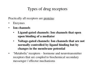
Nicotinic-acetylcholine (nAChR) receptors *cholinergic ligand-gated cationic channels * Most nAChRs are presynaptic neurons *composed of several subunits randomly arranged to give receptors that are slightly different. * subunits (2-7 and 2-4); form pentameric channels that are 300 kDa
Acetylcholine at dentritic portion of neuron is received by nAChR • allows the influx of Ca 2+ • Causes release of synaptic vessicles
Nicotine • agonist • decreases receptor breakdown • inhibits monoamine oxidase B, the enzyme that breaks down dopamine
Stimulation of the nAChR receptors causes an influx in Na+ and Ca2+. • influx depolarizes the cell and regulates the release of neurotransmitters, • modulates neurite outgrowth, • Neurite out growth plays an important role in enhancing cognition and reducing dementia • activation of secondary messengers • Early gene transcription
The different combinations of subunits give rise to the different pharmacologic properties. • 7 subunits have a high permeability to Ca2+ • Combinations of subunits affect the rate of desensitization. • 32 desensitizes 10 times faster than 34 or 44
Protective subunits • 7 has neuro-protective against glutamate toxicity, adverse effects of nerve growth factor deprivation and ethanol induced cytotoxicity. • Activation of 42 containing receptors protects against -amyloid-induced toxicity
Objective To: • Observe the distribution of mRNA subunits in the substantia nigra in a monkey model after inducing Parkinsons using MPTP. • Compare this mRNA distribution to that of the mesencephalic (midbrain) rodent model. • Observe the neuro-protective effect of nicotine in a Parkinsonian mesencephalic rodent model.
Experiment: Monkey Model • Samiri sciureus, Squirrel monkeys weighing 0.7 –1.0 kg • Baseline locomotor activity was measured using computerized movement monitor cage containing infrared sensors. • 2 mg/kg treatment of saline or 1-methyl-4-phenyl-1,2,3,6-tetrahydropteridine (MPTP) subcutaneously.
2 ½ weeks after treatment with MPTP, locomotor activity was again monitored • The monkeys were killed and the effects of MPTP were measured.
Tissue Samples: • Tissue samples were cut into blocks 6mm thick. • Dopamine and Homovanillic acid (HVA) were extracted and measured from the caudate and putamen (from the basal ganglia) using 0.4 N perchloric acid and chromatography.
Results: The effects of MPTP • A decline in motor activity decreased more than 50% when compared to the saline control. • The levels of dopamine and HVA were reduced by 50% when compared to the saline.
Objective: Significance: To investigate the distribution of 4, 6, 7, 2, 3 and 4 mRNA in the Squirrel monkey basal ganglia. To observe the subunits greatly affected during degeneration and nicotine treatment. Distribution of subunit mRNA
4 and 2 are predominant subunits in neuronal nicotinic receptors • 7 primary subunit in bungarotoxin (BGT) nicotinic receptors • 6 and 3 have a unique distribution in mice.
Distribution of subunit mRNA • The blocks were further cut into 20m thick sections to carry out in situ hybridizations • Thawed onto poly-L-lysine coated slides and stored at -80°C.
cDNA of the different subunits found in large cytoplasmic loops were used to sequence cRNA probes. • A transcription Kit was used to carry out transcription. [35S]UTP was used as a label.
Slides with hybridized cRNA were placed against Hyperfilm -max with 14C-radioactive standards. • Development time: 6-8 weeks • The optical and photographic density of the labeled probes were determined using Computer densitometry.
Results: Subunit mRNA after MPTP • The lesions formed were similar to those in found in the early stages of Parkinson’s disease. • There were no changes in 4, 7, 2 and 4 mRNA in the substantia nigra. • 6 mRNA increased and 3 mRNA decreased
Objective: To compare the distribution of receptor subunits using immuno-stains. To observe the effects of MPP+ (a metabolite of MPTP) after nicotine treatment. Mesencephalic culture model
Mesencephalic culture model • Ventral mesencephalon portions of E15 Sprague-Dawley mice. • dissected and the tissue cultures were prepared like the those extracted from the monkeys. • Cut into 6mm thick blocks • Dopamine and HVA extracted
Samples were centrifuged and incubated in 0.5% trypsin-ethylenediaminetetraacetic acid. • Culture medium stop further reactions. • Centrifuged re-suspended in 5ml of the culture media.
Then they were plated on poly-D-Lysine Nunclon 48-well dishes at a density of 3 X 10⁵ cells/cm2. • Added [125I]-BGT (bungarotoxin) was to intact cultures to measure the activity of 7 containing receptors. • Added [3H]Epibatidine to membranes prepared from the culture to measure the activity of 2 and 6 containing receptors
Tyrosine hydroxylase immunochemistry was used to measure the number of dopaminergic neurons. • Special culture media consisting of goat serum, bovine serum albumin, polyvinyl pyrrolidone and Triton-X-100, PBS.
Left for 1 hr at room temperature • Tyrosine hydroxylase was added and left overnight at 4C • Washed and then bound to immunoglobulins
Immuno-stained cells were counted under 100x magnification. • 24 hrs prior to exposure to MPP+, samples were incubated in 10M nicotine. • 3 M of MPP+ (active metabolite of MPTP) was added to dopaminergic neurons and compared to the tissue samples before they were exposed to toxins.
Results: Monkey vs Mouse Distribution of subunit mRNA The distribution of subunit mRNA in the Caudate and Putamen in the monkey model • 4 and 7 nicotinic mRNAs were present • Low density of 2 subunits and high densities of 4 subunits were observed . • 4, 6, 2 and 3 mRNA was found in the rodent mesencephalic culture model.
Results: Monkey vs Mouse Distribution of subunit mRNA • 4, 6, 7, 2, 3 and 4 mRNA was found in the monkey substantia nigra. • 4, 6, 2 and 3 mRNA was found in the rodent mesencephalic culture model. • The distribution of subunits in the mRNA in rodents and monkeys are similar
Results: Mesencephalic culture • The effects of neurotoxicity MPP+ on mesencephalic culture was measured by comparing the number of neurons before and after the application.
Before MPP+ the number of cells immunostained. • [3H]epibatidine 2.2 ± 0.4fmol/106 cells • [125I ]-BGT 0.5 ± 0.1 fmol/106 cells • There was a 80-95% destruction in the nigrostriatal in the rodent mesencephalon model. • After pre-nicotine treatment there was a 20% decrease in dopaminergic cells.
Conclusion • Nicotinic receptors are cholinergic receptors that are made up of subunits that affect the activity of the receptor. • There is some similarity in the distribution of subunits in brain of rodents and monkeys, however, the levels of expression differs. • Locomotor activity decreases with MPTP toxicity.
MPTP and MPP+ toxicity reduces the number of dopaminergic neurons. • Pre-treatment of nicotine reduces the loss of dopaminergic neurons caused by MPP+ • The neuroprotective effects may be due to the activation of receptors containing specific subunits.
References: • Court, J.A., Ruiz-Martin, C., Graham, A., and Elaine Perry. (2000) Nicotinic receptors in human brain: topography and pathology. Journal of Chemical Neuroanatomy Vol 20: 281-298 • Mihailescu, Stefan Drucker-Colin, Rene (2000) Nicotine, brain nicotinic receptors and neuropsychiatric disorders. Archives of Medical Research Vol 31: 131-144 • Bondy, S., Ali, S.F., and M. Kleinman (2000) Exposure of mice to tobacco smoke attenuates the toxic effect of methamphetamine on dopamine systems. Toxicology Letters Vol 118: 43-46 • Costa, G., Abin-Carriquiry, J.A. and Federico Dajas. (2001) Nicotine prevents striatial dopamine loss produced by 6-hydroxydopamine lesion in the substantia nigra. Brain Research Interactive Vol 888: 336-342. • Encyclopedia Britannica. www.britanica.com • Quik, M. and G. Jeyarasasingam (2000) Nicotinic receptors and parkinson’s disease. European Journal of Pharmacology Vol 393: 223-230.