Download
1 / 22
250 likes | 470 Views
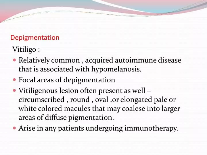
Depigmentation. Vitiligo : Relatively common , acquired autoimmune disease that is associated with hypomelanosis. Focal areas of depigmentation

E N D
Depigmentation Vitiligo : • Relatively common , acquired autoimmune disease that is associated with hypomelanosis. • Focal areas of depigmentation • Vitiligenous lesion often present as well – circumscribed , round , oval ,or elongated pale or white colored macules that may coalese into larger areas of diffuse pigmentation. • Arise in any patients undergoing immunotherapy.
Hypomelanosis of inner & outer surfaces of lips & perioral skin may be seen in up to 20% of patients. • Treatment with tropical corticosteroids , systemic photochemical therapies (psoralen & ultraviolet A exposure proven effective. • Medicinal depigmentation that is cutaneoua bleaching to create unified skin color. • Surgical intervention may be only optional (autologus epithelial grafts) used succesfully.
HEMOGLOBIN AND IRON ASSOCIATED PIGMENTATION • Ecchymosis: • Traumatic ecchymosis is common on the lips and face yet is uncommon in oral mucosa except in cases of blunt force trauma and oral intubation. • Immediately following traumatic event erythrocyte extra vasation into the sub mucosa will appear as bright red macule. • The lesion will assume brown coloration within few days, after hemoglobin is degraded to hemosiderin.
PURPURA/PETECHIAE • Petechiae typically characterised as being pinpoint or slightly larger than pinpoint and purpura as multiple, small 2 to 4 mm collections of extravasated blood. • Oral purpura may develop as a consequence of trauma or viral or systemic disease, identified in soft palate although any mucosal site may be affected.
HEMOCHROMATOSIS • Chronic, progressive disease that is characterized by excessive iron deposits (usually in the form of hemosiderin) in the liver and other organs and tissues. • Oral mucosal pigmentation is also well reorganized. • Oral pigmentation is often diffuse and brown to gray in appearance. • Palate and gingiva are most commonly affected.
EXOGENOUS PIGMENTATION • AMALGUM TATTO: • Etiology is deposition of amalgam material into sub mucosal tissue. • Lesions small, asymptomatic, macular and bluish gray or oven black in appearance. • Gingiva, alveolar mucosa, buccal mucosa and floor of mouth are most common sites. • Lesions found in vicinity of teeth with large amalgam restoration or crowned teeth and also in around healed extraction sites.
GRAPHITE TATTOO • Commonly seen on palate and represent traumatic implantation of graphite particles from a pencil • Lesion present similar to amalgam tattoo. So biopsy is often warranted.
ORNAMENTAL TATTOOS • South African female tribal custom includes brushing the teeth and gums with a chewed root of the tree Euclea natalensis with the belief that promotes oral health. • Plant root contain napthoquinones are pigmented and the mouths of root users are typically bright orange.
MEDICINAL METAL INDUCED PIGMENTATION • Silver may cause generalized blue-gray discoloration (argyria). • Gold induced pigment may appear blue-gray or purple (chrysiasis). • Generalized black pigmentation of tongue due to chewing of bismuth sub salicylate tablets, a commonly used antacid.
HEAVY METAL PIGMENTATION • Lead, mercury, bismuth and arsenic have shown to be deposited in oral tissue if ingested in sufficient quantities over a extended period of time. • Found along the free marginal gingiva, metallic line usually gray-black appearance. • Signs and symptoms of metal poisoning neurologic disorders, intestinal pain and sialorrhea.
HAIRY TONGUE • Change in oral flora associated with chromic antibiotic therapy. • Colonization of chromogenic bacteria impart variety of colors white, green, brown or black. • Various foods, drinks and also smoking of tobacco or crack cocaine has been shown in black hairy tongue. • Treatment is using tongue scrapper and limit ingestion of coloring foods.