Download
1 / 63
780 likes | 1.66k Views
Meninges, CSF & Ventricular system. Objectives. Describe the arrangement of the meninges and their relationship to brain and spinal cord. Explain the occurrence of epidural, subdural and subarachnoid spaces. Locate the principal subarachnoid cisterns, and arachnoid granulations.

E N D
Objectives • Describe the arrangement of the meninges and their relationship to brain and spinal cord. • Explain the occurrence of epidural, subdural and subarachnoid spaces. • Locate the principal subarachnoid cisterns, and arachnoid granulations. • Describe the ventricles of brain and importance of their choroids plexus. • Summarize the pathway of cerebrospinal fluid (CSF) circulation • Identify brain ventricles in CT scan, MRI .
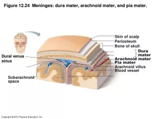
MENINGES • The brain and spinal cord are invested by three concentric membranes ; • The outermost layer is the dura matter. • The middle layer is the arachnoid matter. • The innermost layer is the pia matter.
Cranial Meninges - 3 layer protective membrane • 1.Dura Mater- Composed of two layers • a) Periosteal– outer layer, attaches to bone. • b) Meningeal– inner layer, closer to brain. • Two layers fused, except to enclose the dural sinuses • 2.Arachnoid Layer- ‘spider’ web like. • 3. Pia Mater - delicate, follows convolutions.
Cranial Meningeal Spaces Epidural space Potential space superior to dura. Subdural space Potential space between dura and arachnoid mater. Subarachnoid space Filled with CSF Contains the blood vessels supplying brain.
In the cranial vault, the epidural space is normally closed since the dura is attached to the periosteum which is attached to the inner surface of the skull In the spinal cord the epidural space is filled with adipose tissue.
DURA MATER The cranial durais a two layered tough, fibrous membrane that surrounds the brain. It is formed of two layers;periosteal and meningeal. The periosteal layer is attached to the skull. The meningeal layer is folded forming the dural folds; falxcerebri, and tentoriamcerebelli. Sensory innervation of the dura is mostly from the three branches of the trigeminal and vagus nerves & C1 to C3.
DURA MATER Two large reflection of dura extend into the cranial cavity 1.The falxcerebri, In the midline, It is a vertical sickle shaped sheet of dura, extends from the cranial roof into the great longitudinal fissure between the two cerebral hemispheres. It has an attached border adherent to the skull. And a free border lies above the corpus callosum.
DURAMATER 2. A horizontal shelf of dura, The tentoriumcerebelli, It lies between the posterior part of the cerebral hemispheres and the cerebellum. It has a free border that encircles the midbrain. In the middle line it is continuous with the falxcerebri,
Arachnoid Mater& Pia Mater The arachnoid mater is a soft, translucent membrane loosely envelops the brain. The arachnoid mater is separated from the dura by a narrow subduralspace. The pia mater is the innermost, thin, delicate & highly vascular membrane that is closely adherent to the gyri and fitted into the sulci. Between the pia and arachnoid mater lies the subarachnoid space which contains; fibrous trabeculae, main blood vessels and CSF.
Subarachnoid Space It is varied in depth forming; subarachnoid cisterns. The cisterna magna, or cerebllomedullary cistern which lies between the inferior surface of the cerebellum and the back of the medulla. At this cistern CSF flows out of the fourth ventricle.
Subarachnoid Space The interpeduncular cistern, which is located at the base of the brain, where the arachnoid spans the space between the two cerebral peduncles. This cistern contains the optic chiasma & circulusarteriosus of Willis.
Subarachnoid Space • Pontine cistern(Prepontinecistern or cisterna pontis). Surrounds the ventral aspect of the pons. It contains: • The basilar artery and the origin of the anteroinferior cerebellar artery. • The origin of the superior cerebellar arteries. • The sixth (VI) cranial nerve
CSF • CSF is clear, colorless, and odorless fluid • produced within the ventricles secreted by the Choroid plexus.Choroid plexuses are areas where the lining wall of the ventricle is very thin and has a profusion of capillaries • provides mechanical support – protection from pressure changes. • In adults, the total volume of CSF is about 150 ml • Between 400 and 500 mL of CSF is produced and reabsorbed daily. • Thus the CSF turns over about 3.7 times a day. This continuous flow into the venous system dilutes the concentration of larger, lipoid soluble molecules penetrating the brain and CSF.
Choroid Plexus • The lining ependyma of each ventricle comes into contact with the surface pia mater allowing the invagination of a mass of blood capillaries --- combination of these capillaries, pia and ependymaconstitutes the choroid plexus.
Pia and Ependyma Pia Ependyma Diagrammatic Cross Section The “hollow” brain
Pia and Ependyma In some locations pia mater and ependyma come together. A plexus of blood vessels “invaginates” the thin layer. Cerebrospinal fluid enters the ventricle across this membrane.
CSF Formation • The cerebrospinal fluid is formed mainly in the choroid plexuses of the lateral, third, and fourth ventricles; • Some originate from the ependymal cells lining the ventricles and from the brain substance through the perivascular spaces.
Choroid Plexus • lateral ventricles • continuous through Interventricular foramen with the small plexus in the third ventricle. • secretes the bulk of the CSF • fourth ventricle • separate from that in the third and lateral ventricles • only makes a small contribution to the total amount of CSF
Circulation • The circulation begins with its secretion from the choroid plexuses in the ventricles • The fluid passes from the lateral ventricles into the third ventricle through the interventricular foramina • It then passes into the fourth ventricle through the narrow cerebral aqueduct. • The circulation is aided by the arterial pulsations of the choroid plexuses and by the cilia on the ependymal cells lining the ventricles. • From the fourth ventricle, the fluid passes slowly through the median aperture and the lateral foramina of the lateral recesses of the fourth ventricle and enters the subarachnoid space
CSF Drainage • The CSF returns to the vascular system by entering the dural venous sinusesvia the arachnoid granulations(or villi). • The arachnoid villi act as one-way valves between the subarachnoid space and the dural sinuses. The rate of absorption correlates with the CSF pressure.
CSF Drainage • Flows along the cranial nervesand spinal nerve roots into the lymphatic channels; this flow may play a substantial role in CSF reabsorption, in particular in the neonate, in which arachnoid granulations are sparsely distributed.
Summary • Filtered from blood in: • Choroid plexuses • Circulates in through ventricles, canals, & between meninges • Returned to blood at superior sagittal sinus mainly in addition to other venous sinuses • Along nerves to extra cranial lymphatics • Through venous plexuses in vertebral canal and around spinal cord
VENTRICULAR SYSTEM • Comprises of: • two lateral ventricles • third ventricle • cerebral aqueduct • the fourth ventricle
VENTRICULAR SYSTEMfrom the rightfrom above Central canal
The Lateral Ventricle • Is the cavity inside the cerebral hemisphere • The 2 lateral ventricles are the largest of the ventricles. • They are irregular in shape. • Each consists of a central part, with anterior, posterior and inferior horns.
Lateral ventricles • C-shaped cavity • within each cerebral hemisphere • Consists of: • anterior horn - frontal lobe • body --- parietal lobe • posterior horn ---- occipital lobe • inferior horn ---- temporal lobe
Anterior horn (Frontal lobe) Is anterior to the interventricularforamen(of Monro) . Its roof and anterior border are formed by the corpus callosum, Its vertical medial wall by the septum pellucidum. The floor is formed by the head of the caudate nucleus.
Central part • Triangular in shape, having : • Roof: corpus callosum; • Medial wall: by the posterior part of the septum pellucidum • Floor and inferolateral wall:by parts of the caudate nucleus, thalamus, choroid plexus and fornix.
Thalamus Thalamus and Basal ganglia Brain stem
Body of Caudate nucleus Lentiform nucleus Head of Caudate nucleus Thalamus Amygdaloid body Tail of Caudate nucleus
Posterior Horn • Extends into the occipital lobe. • Its roof is formed by fibers of the corpus callosum. • It is the most variably developed and may even be absent.
Callosal radiation Cingulate sulcus Optic radiation Posterior horn of lateral ventricle Collateral eminence Collateral sulcus Splenium of corpus callosum Tapetum of corpus callosum
Inferior Horn • It traverses the temporal lobe. • Its roof is formed by the white substance of the cerebral hemisphere. • Along the medial border is the striaterminalis and the tail of the caudate nucleus. • The floor and the medial wall are formed by the fimbria, the hippocampus and the collateral eminence. • The amygdaloid nucleus bulges into the terminal part of the inferior horn.
Third Ventricle • It is a narrow cavity between the 2 thalami. • is a narrow vertical cleft between the 2 lateral ventricles • It communicates with the 2 lateral ventricles through the interventricular foramen and with the 4th ventricle through the cerebral aquiduct. • Comprises of: • Anterior wall • Two side walls • Floor • Roof
Third Ventricle • Anterior wall: • lamina terminalis • anterior commissure • Roof: The roof is formed by the telachoroidea • Floor: • optic chiasma • tuber cinereummedian eminence • infundibulum • mamillary bodies • posterior perforated substance • tegmentum of the cerebral peduncles • Two side walls: • Thalamus Interthalamic adhesion (60% of brains) • Hypothalamus Supraoptic nucleus – ADH Paraventricular nucleus – Vasopressin/Oxytocin • SubthalamusSubthalamic nucleus
Telachoroidea Telachoroidea (double fold of pia) • Reflection of two layers of pia matter • Medially b/w interventricular foramina • Laterally across the upper surface of thalamus