Download
1 / 32
460 likes | 1.11k Views
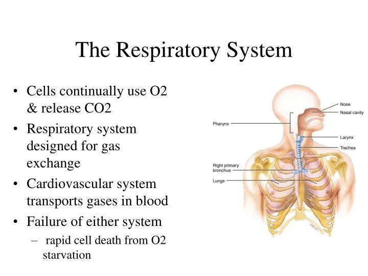
The Respiratory System. Cells continually use O2 & release CO2 Respiratory system designed for gas exchange Cardiovascular system transports gases in blood Failure of either system rapid cell death from O2 starvation. Human Lungs. Respiratory System Anatomy. Nose Pharynx = throat

E N D
The Respiratory System • Cells continually use O2 & release CO2 • Respiratory system designed for gas exchange • Cardiovascular system transports gases in blood • Failure of either system • rapid cell death from O2 starvation
Respiratory System Anatomy • Nose • Pharynx = throat • Larynx = voicebox • Trachea = windpipe • Bronchi = airways • Lungs • Locations of infections • upper respiratory tract is above vocal cords • lower respiratory tract is below vocal cords
Trachea • Size is 5 in long & 1in diameter • Extends from larynx to T5 anterior to the esophagus and then splits into bronchi • Layers • mucosa = pseudostratified columnar with cilia & goblet • submucosa = loose connective tissue & seromucous glands • hyaline cartilage = 16 to 20 incomplete rings • open side facing esophagus contains trachealis m. (smooth) • internal ridge on last ring called carina (cough reflex) • adventitia binds it to other organs
Histology of the Trachea • Ciliated pseudostratified columnar epithelium • Hyaline cartilage as C-shaped structure closed by trachealis muscle
Airway Epithelium • Ciliated pseudostratified columnar epithelium with goblet cells produce a moving mass of mucus.
Bronchi and Bronchioles • Primary bronchi supply each lung • Secondary bronchi supply each lobe of the lungs (3 right + 2 left) • Tertiary bronchi supply each bronchopulmonary segment • Repeated branchings called bronchioles form a bronchial tree
Histology of Bronchial Tree • Epithelium changes from pseudostratified ciliated columnar to nonciliated simple cuboidal, and finally to simple squamous as pass deeper into lungs • Incomplete rings of cartilage replaced by rings of smooth muscle & then connective tissue • sympathetic NS & adrenal gland release epinephrine that relaxes smooth muscle & dilates airways • asthma attack or allergic reactions constrict distal bronchiole smooth muscle
Structures within a Lobule of Lung • Branchings of single arteriole, venule & bronchiole are wrapped by elastic CT • Respiratory bronchiole • simple squamous • Alveolar ducts surrounded by alveolar sacs & alveoli • sac is 2 or more alveoli sharing a common opening
Histology of Lung Tissue Photomicrograph of lung tissue showing bronchioles, alveoli and alveolar ducts.
Cells Types of the Alveoli • Type I alveolar cells • simple squamous cells where gas exchange occurs • Type II alveolar cells (septal cells) • free surface has microvilli • secrete alveolar fluid containing surfactant • Alveolar dust cells • wandering macrophages remove debris
Alveolar-Capillary Membrane • Respiratory membrane = 1/2 micron thick • Exchange of gas from alveoli to blood • 4 Layers of membrane to cross • alveolar epithelial wall of type I cells • alveolar epithelial basement membrane • capillary basement membrane • endothelial cells of capillary • Vast surface area = handball court
Double Blood Supply to the Lungs • Deoxygenated blood arrives through pulmonary trunk from the right ventricle • Bronchial arteries branch off of the aorta to supply oxygenated blood to lung tissue • Venous drainage returns all blood to heart
Breathing or Pulmonary Ventilation • Air moves into lungs when pressure inside lungs is less than atmospheric pressure • Increase volume of lungs, Boyle’s law • Air moves out of the lungs when pressure inside lungs is greater than atmospheric pressure • Passive process • Atmospheric pressure = 1 atm or 760mm Hg
Boyle’s Law • As the size of closed container decreases, pressure inside is increased, inverse relationship • The molecules have less wall area to strike so the pressure on each inch of area increases.
Dimensions of the Chest Cavity • Breathing in requires muscular activity & chest size changes • Contraction of the diaphragm flattens the dome and increases the vertical dimension of the chest • Contraction of external intercostal muscles elevates ribs and increases diameter of chest
Inspiration • Diaphragm moves 1 cm & ribs lifted by muscles • Intrathoracic pressure falls and 2-3 liters inhaled
Expiration • Passive process with no muscle action • Elastic recoil & surface tension in alveoli pulls inward • Alveolar pressure increases & air is pushed out
Summary of Breathing • Alveolar pressure decreases & air rushes in • Alveolar pressure increases & air rushes out
Alveolar Surface Tension • Thin layer of fluid in alveoli causes inwardly directed force = surface tension • water molecules strongly attracted to each other • Causes alveoli to remain as small as possible • Detergent-like substance called surfactant produced by Type II alveolar cells • lowers alveolar surface tension • insufficient in premature babies so that alveoli collapse at end of each exhalation
Pneumothorax • Pleural cavities are sealed cavities not open to the outside • Injuries to the chest wall that let air enter the intrapleural space • causes a pneumothorax • collapsed lung on same side as injury • surface tension and recoil of elastic fibers causes the lung to collapse
Lung Volumes and Capacities • Tidal volume = amount air moved during quiet breathing • MVR= minute ventilation is amount of air moved in a minute • Reserve volumes ---- amount you can breathe either in or out above that amount of tidal volume • Residual volume = 1200 mL permanently trapped air in system • Vital capacity & total lung capacity are sums of the other volumes
Dalton’s Law • Each gas in a mixture of gases exerts its own pressure • as if all other gases were not present • partial pressures denoted as p • Total pressure is sum of all partial pressures • atmospheric pressure (760 mm Hg) = pO2 + pCO2 + pN2 + pH2O
External Respiration • Gases diffuse from areas of high partial pressure to areas of low partial pressure • Exchange of gas between air & blood • Deoxygenated blood becomes saturated • Compare gas movements in pulmonary capillaries to tissue capillaries
Internal Respiration • Exchange of gases between blood & tissues • Conversion of oxygenated blood into deoxygenated • Observe diffusion of O2 inward • at rest 25% of available O2 enters cells • during exercise more O2 is absorbed • Observe diffusion of CO2 outward
Hemoglobin 4 heme molecules each bind one oxygen molecule
Oxygen Transport in the Blood • Oxyhemoglobin contains 98.5% chemically combined oxygen and hemoglobin • inside red blood cells • Does not dissolve easily in water • only 1.5% transported dissolved in blood • Only the dissolved O2 can diffuse into tissues • Factors affecting dissociation of O2 from hemoglobin are important
Carbon Dioxide Transport • 100 ml of blood carries 55 ml of CO2 • Is carried by the blood in 3 ways • dissolved in plasma • combined with the globin part of Hb molecule forming carbaminohemoglobin • as part of bicarbonate ion • CO2 + H2O combine to form carbonic acid that dissociates into H+ and bicarbonate ion
Smokers Lowered Respiratory Efficiency • Smoker is easily “winded” with moderate exercise • nicotine constricts terminal bronchioles • carbon monoxide in smoke binds to hemoglobin • irritants in smoke cause excess mucus secretion • irritants inhibit movements of cilia • in time destroys elastic fibers in lungs & leads to emphysema • trapping of air in alveoli & reduced gas exchange


