Download
1 / 1
30 likes | 172 Views
Mice model of central post stroke pain. Hsi-Chien Shih, Bai -Chung Shyu . Institute of Biomedical Sciences, Academia Sinica , Taipei, Taiwan. Abstract

E N D
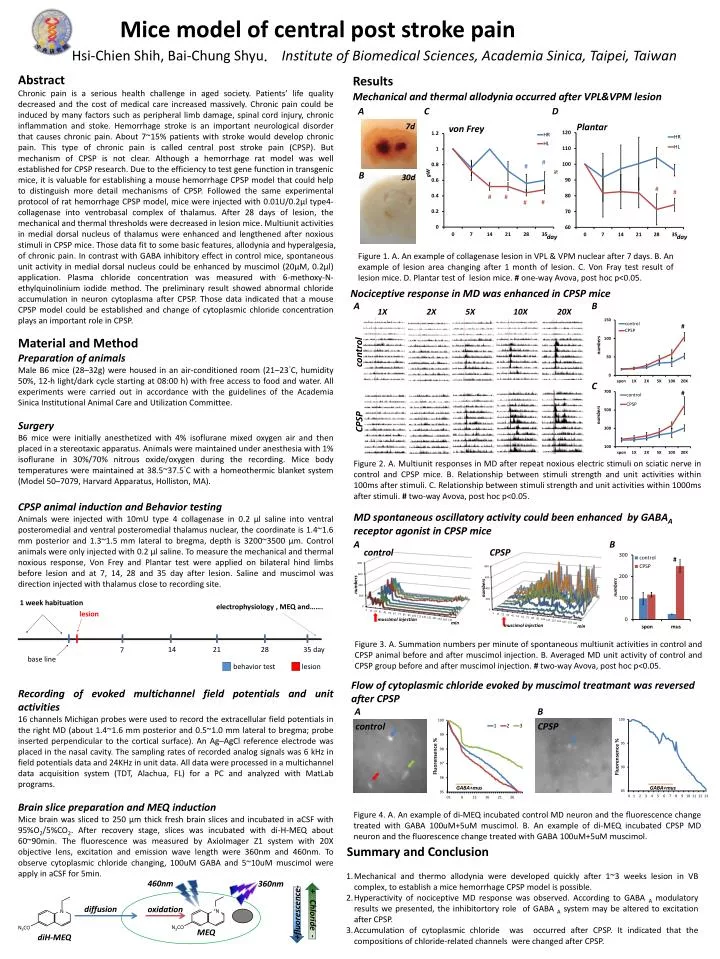
Mice model of central post stroke pain Hsi-ChienShih, Bai-Chung Shyu.Institute of Biomedical Sciences, Academia Sinica, Taipei, Taiwan Abstract Chronic pain is a serious health challenge in aged society. Patients’ life quality decreased and the cost of medical care increased massively. Chronic pain could be induced by many factors such as peripheral limb damage, spinal cord injury, chronic inflammation and stoke. Hemorrhage stroke is an important neurological disorder that causes chronic pain. About 7~15% patients with stroke would develop chronic pain. This type of chronic pain is called central post stroke pain (CPSP). But mechanism of CPSP is not clear. Although a hemorrhage rat model was well established for CPSP research. Due to the efficiency to test gene function in transgenic mice, it is valuable for establishing a mouse hemorrhage CPSP model that could help to distinguish more detail mechanisms of CPSP. Followed the same experimental protocol of rat hemorrhage CPSP model, mice were injected with 0.01U/0.2μl type4-collagenase into ventrobasal complex of thalamus. After 28 days of lesion, the mechanical and thermal thresholds were decreased in lesion mice. Multiunit activities in medial dorsal nucleus of thalamus were enhanced and lengthened after noxious stimuli in CPSP mice. Those data fit to some basic features, allodynia and hyperalgesia, of chronic pain. In contrast with GABA inhibitory effect in control mice, spontaneous unit activity in medial dorsal nucleus could be enhanced by muscimol (20μM, 0.2μl) application. Plasma chloride concentration was measured with 6-methoxy-N-ethylquinolinium iodide method. The preliminary result showed abnormal chloride accumulation in neuron cytoplasma after CPSP. Those data indicated that a mouse CPSP model could be established and change of cytoplasmic chloride concentration plays an important role in CPSP. Results Mechanical and thermal allodynia occurred after VPL&VPM lesion A C D 7d Plantar von Frey # # % gW B 30d # # # # # # day day Figure 1. A. An example of collagenase lesion in VPL & VPM nuclear after 7 days. B. An example of lesion area changing after 1 month of lesion. C. Von Fray test result of lesion mice. D. Plantar test of lesion mice. # one-way Avova, post hoc p<0.05. Nociceptiveresponse in MD was enhanced in CPSP mice A B 1X 2X 5X 10X 20X # Material and Method Preparation of animals Male B6 mice (28–32g) were housed in an air-conditioned room (21–23°C, humidity 50%, 12-h light/dark cycle starting at 08:00 h) with free access to food and water. All experiments were carried out in accordance with the guidelines of the Academia Sinica Institutional Animal Care and Utilization Committee. Surgery B6 mice were initially anesthetized with 4% isoflurane mixed oxygen air and then placed in a stereotaxic apparatus. Animals were maintained under anesthesia with 1% isoflurane in 30%/70% nitrous oxide/oxygen during the recording. Mice body temperatures were maintained at 38.5~37.5°C with a homeothermic blanket system (Model 50–7079, Harvard Apparatus, Holliston, MA). CPSP animal induction and Behavior testing Animals were injected with 10mU type 4 collagenase in 0.2 μl saline into ventral posteromedial and ventral posteromedial thalamus nuclear, the coordinate is 1.4~1.6 mm posterior and 1.3~1.5 mm lateral to bregma, depth is 3200~3500 μm. Control animals were only injected with 0.2 μl saline. To measure the mechanical and thermal noxious response, Von Frey and Plantar test were applied on bilateral hind limbs before lesion and at 7, 14, 28 and 35 day after lesion. Saline and muscimol was direction injected with thalamus close to recording site. Recording of evoked multichannel field potentials and unit activities 16 channels Michigan probes were used to record the extracellular field potentials in the right MD (about 1.4~1.6 mm posterior and 0.5~1.0 mm lateral to bregma; probe inserted perpendicular to the cortical surface). An Ag–AgCl reference electrode was placed in the nasal cavity. The sampling rates of recorded analog signals was 6 kHz in field potentials data and 24KHz in unit data. All data were processed in a multichannel data acquisition system (TDT, Alachua, FL) for a PC and analyzed with MatLab programs. Brain slice preparation and MEQ induction Mice brain was sliced to 250 μm thick fresh brain slices and incubated in aCSF with 95%O2/5%CO2. After recovery stage, slices was incubated with di-H-MEQ about 60~90min. The fluorescence was measured by AxioImager Z1 system with 20X objective lens, excitation and emission wave length were 360nm and 460nm. To observe cytoplasmic chloride changing, 100uM GABA and 5~10uM muscimol were apply in aCSF for 5min. numbers control C # numbers CPSP Figure 2. A. Multiunit responses in MD after repeat noxious electric stimuli on sciatic nerve in control and CPSP mice. B. Relationship between stimuli strength and unit activities within 100ms after stimuli. C. Relationship between stimuli strength and unit activities within 1000ms after stimuli. #two-way Avova, post hoc p<0.05. MD spontaneous oscillatory activity could been enhanced by GABAAreceptor agonist in CPSP mice A B control CPSP # numbers numbers numbers 1 week habituation electrophysiology , MEQ and……. lesion muscimol injection min muscimol injection min Figure 3. A. Summation numbers per minute of spontaneous multiunit activities in control and CPSP animal before and after muscimol injection. B. Averaged MD unit activity of control and CPSP group before and after muscimolinjection. # two-way Avova, post hoc p<0.05. 14 21 28 35 day 7 base line behavior test lesion Flow of cytoplasmic chloride evoked by muscimoltreatmant was reversed after CPSP A B control CPSP Fluorensence % Fluorensence % GABA+mus GABA+mus Figure 4. A. An example of di-MEQ incubated control MD neuron and the fluorescence change treated with GABA 100uM+5uM muscimol. B. An example of di-MEQ incubated CPSP MD neuron and the fluorescence change treated with GABA 100uM+5uM muscimol. Summary and Conclusion • Mechanical and thermo allodynia were developed quickly after 1~3 weeks lesion in VB complex, to establish a mice hemorrhage CPSP model is possible. • Hyperactivity of nociceptive MD response was observed. According to GABA Amodulatory results we presented, the inhibitortory role of GABA A system may be altered to excitation after CPSP. • Accumulation of cytoplasmic chloride was occurred after CPSP. It indicated that the compositions of chloride-related channels were changed after CPSP. 460nm 360nm N +fluorescence- diffusion oxidation + Chloride - N3CO +N N3CO MEQ diH-MEQ