Download
1 / 79
790 likes | 802 Views
Structure determination by NMR. NMR principles Data acquisition Spectra process xwinnmr 、 nmrpipe 、 nmrview 、 Topspin Assignment sparky Data Analysis Structure determination InsightII 、 Xplor 、 CNS Structural analysis Procheck 、 Molmol 、 Pymol. ~~NMR Experiments studies~~.

E N D
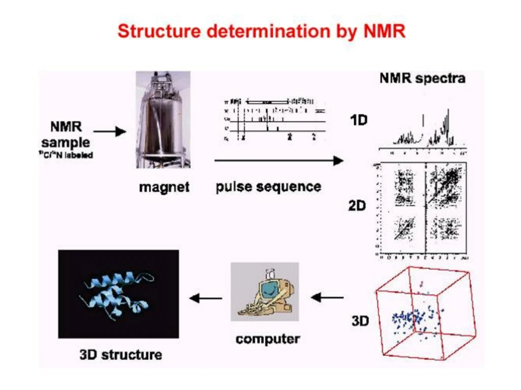
Structure determination by NMR • NMR principles • Data acquisition • Spectra process • xwinnmr、nmrpipe、nmrview、Topspin • Assignment • sparky • Data Analysis • Structure determination • InsightII、Xplor、CNS • Structural analysis • Procheck、Molmol、Pymol
Sample prepare • High concentrated protein • 10mg-30mg • Proton labeling • H1 • H1-N15 • H1-N15-C13 • Limitation • Protein molecular size <25 Kda
Modern Fourier transform NMR spectrometer Coil and superconductor LN2 and LHe2 tank
Spectra process and Assignment • Chemical shifts in proteins
Spectra process and Assignment • Chemical shifts in proteins the a-proton is always around 4 ppm; the aromatic protons are around 7 ppm; the backbone amides at 8 ppm.
Hα,H2O CH,CH2,CH3 HN, aromatic Well-dispersed 1D Spectrum
Why do we go beyond one dimension? • To resolve the crowded signals in 1D spectrum by spreading them into other dimensions. • To elucidate the “through-bond” and “through-space” relationships between the spins in the molecules.
The functions of the different periods of a two-dimensional NMRexperiment are summarized as follows: • Preparation: The desired nonequilibrium state of the spin system is prepared from the initial (equilibrium) state of the spin system. The preparation period in its simplest form consists of a single pulse that generates transverse magnetization, but more complex sequences of pulses can be used to prepare other coherences, such as multiple quantum coherences, and to perform solvent suppression. • Evolution: The off-diagonal components of the density operator prepared in step (1) evolve under the Hamiltonian, He . During the course of the experiment, the incrementable time t1 normally begins at an initial value and increases in discrete steps to a maximum value, t1max. The Hamiltonian, He , may be the free-precession Hamiltonian or may include applied rf fields. The frequencies with which the detected coherence evolves during t1 results in signals appearing at those frequencies in the F1 dimension of the final two-dimensional spectrum. This process is known as F1 frequency labeling of the coherence. • Mixing: During the mixing period, coherence is transferred from one spin to another. The mixing period is the key to establishing the type of correlation between the two dimensions and consequently dictates the information content of the spectrum. Depending on the type of experiment, the mixing period consists of one or more pulses and delays. • Acquisition: The FID is recorded in the conventional fashion. As discussed in Section 4.3, if more than one coherence transfer pathway is feasible, phase cycling or field gradient pulses are used to determine which coherence transfer processes contribute to the final spectrum.
Schematic generation of a three-dimensional NMR experimentfrom the combination of two two-dimensional NMR experiments.
The development of a three-dimensional data set from a two-dimensional data set.
COSY (correlation spectroscopy) • The original 2D experiment. Used to identify nuclei that share a scalar (J) coupling. The presence of off-diagonal peaks (cross-peaks) in the spectrum directly correlates the coupled partners. • NOESY (Nuclear Overhauser Effect Spectroscopy) • A 2D method used to map NOE correlations between protons within a molecule. The spectra have a layout similar to COSY but cross peaks now indicate NOEs between the correlated protons.
The five regions of the COSY spectrum containing the fingerprint cross-peaks.
TOCSY Total Correlation Spectroscopy HOHAHA(homonuclear Hartmann–Hahn) spectroscopy
Pulse sequence and coherence level diagram for the TOCSY experiment.
Sections of H2O TOCSY spectra acquired with mixing times of 48 (left), 83 (center), and 102 ms (right).
Cross-Relaxation NMR Experiments NOESY (Nuclear Overhauser Effect Spectroscopy)
Pulse sequence and coherence level diagram for the NOESY experiment.
Secondary structure elements have characteristic NOE patterns
Assignment Sequential assignment Side chain assignment
Spectra process and Assignment • 2D NMR spectroscopy
Spectra process and Assignment • 2D NMR spectroscopy • 2D TOCSY
Spectra process and Assignment • 2D NMR spectroscopy • 2D NOESY and TOCSY
Spectra process and Assignment • Assignment
Spectra process and Assignment • Assignment – TOCSY : identify spin system HN92 HN91 HN93 0 g b b b 4 0 a Ha91 a Ha93 Ha92 a 10 6 10 0 10 7
Spectra process and Assignment • Assignment - sequential assignment
Spectra process and Assignment • Assignment – NOESY : sequential assignment
Spectra process and Assignment • Assignment - sequential assignment
TOCSY : Amide to Aliphatic Region N’-ACGSC RKKCK GSGKC INGRC KCY-C’
H H H O N C C N C C N H O H NOESY and TOCSY : Amide to a Region
heteronuclear multiple-quantum coherence (HMQC) Heteronuclear single-quantum coherence (HSQC) TROSY
Isotope-labeling of proteins (I)15N labeling • Grow proteins on minimal media (M9) with 15NH4Cl as the sole nitrogen source. • $100-$1000 for mM sample. • Structure elucidation of medium-sized proteins (50-100 a.a.)
Isotope-labeling of proteins (II)15N, 13C labeling • Grow proteins on minimal media (M9) with 15NH4Cl as the sole nitrogen source and 13C-glucose as the sole carbon source. • $1000-$10000 for mM sample. • Structure elucidation of larger proteins (100-250 a.a.)
Isotope-labeling of proteins (III)15N, 13C, 2H labeling • Grow proteins on minimal media (M9) with 15N2H4Cl as the sole nitrogen source and 13C,2H-glucose as the sole carbon source in deuterated water. • Re-exchange deuterium on amide nitrogen to protons. • Strain must be adapted to grow on D2O. • > $10000 for mM sample. • Structure elucidation of larger proteins (> 200 a.a.)
Isotope-labeling of proteins (IV)Site-specific labeling • Add labeled amino acids to non-labeled media. • Assuming that the amino acid is not metabolized, all residues corresponding to that amino acid will be labeled in the protein. • Technique is interesting when structural or dynamic information is only required for specific residues. Thereby, the complete assignment of the protein may be circumvented.