Download

1 / 43
430 likes | 471 Views
Protein Purification. BL4010 10.19.06. Resources. Protein purification: A practical approach. (Harris& Angal IRL Press) Protein purification: Design and scale-up of downstream processing. (Wheelwright Hanser Press)

E N D
Protein Purification BL4010 10.19.06
Resources • Protein purification: A practical approach. (Harris& Angal IRL Press) • Protein purification: Design and scale-up of downstream processing. (Wheelwright Hanser Press) • Methods in Enzymology - several volumes are concerned exclusively with protein purification. • Note that whatever book you get, it is already likely to be out of date.
Why purify? in vitro vs. in vivo analysis
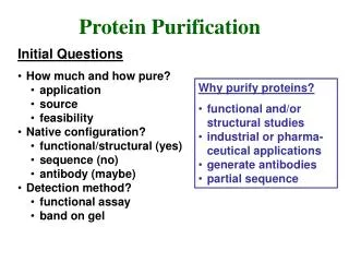
Why purify? • By purifying a protein it can be clearly established that a particular biological activity (enzymatic activity, signaling capacity, etc.) actually resides in a unique protein. • Purified proteins serve as extremely valuable biochemical reagents • Determine mechanism (controlled, observable environment) • Structural determination • Sequence determination • Antibody production • Structure/function analysis - genetic engineering • Finding inhibitors • Detailed kinetic studies
Concentration (size) precipitation ultrafiltration dialysis centrifugation Chromatography (size/charge/chemistry) ion exchange size exclusion affinity Electrophoresis (size/charge) "native" denaturing isoelectric focusing 2-dimensional Immunological (size/charge/chemistry) chromatography in situ imaging immunoblotting The basic techniques
Getting started • Assay (measurable quality) must be specific and convenient • measuring a change in absorbency as NADPH is oxidized in a coupled reaction, • binding activity • a shift of a labeled molecule (DNA, protein) on a gel • the transformation of substrate • the ability to stimulate cell reaction (e.g. proliferation) • Source • easier to purify from a rich source vs. a poor source
Protein Purification Principles • Define objectives • for purity, activity and quantity required of final product to avoid over or under developing a method • Define properties of target protein and critical impurities • to simplify technique selection and optimisation • Develop analytical assays • for fast detection of protein activity/recovery and to work efficiently • Remove damaging contaminants early • for example, proteases
Protein Purification Principles • Use a different technique at each step • to take advantage of sample characteristics which can be used for separation (size, charge, hydrophobicity, ligand specificity) • Minimize sample handling at every stage • to avoid lengthy procedures which risk losing activity/reducing recovery • Minimize use of additives • additives may need to be removed in an extra purification step or may interfere with activity assays • Minimize number of steps - KEEP IT SIMPLE! • extra steps reduce yield and increase time, combine steps logically
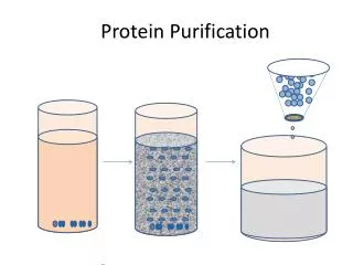
Starting materials • Natural source or artificial expression system • Host for expression, • Bacteria, yeast, plants, transgenic animals • Abundance, contaminants • Lysis and clarification procedures • Native or denaturing conditions • Subcellular fractionation • Selective precipitation • PEI, Streptomycin Sulfate, CTAB for RNA/DNA • Ammonium Sulfate for Proteins
Capture • Quickly remove most damaging contaminants • Concentrate, adsorption methods • Ion Exchange most general • Affinity chromatography can combine capture, intermediate and polishing steps • This step should remove most unwanted contaminants
Intermediate purification • Use a different technique • Affinity chromatography, Hydrophobic interaction chromatography • Starting conditions are specific for each technique • Buffer must be compatible with adsorption • Can change buffer by dialysis or desalting by GFC • Adsorption techniques result in small volume concentrated sample
Polishing • Final removal of trace contaminants • Often size exclusion chromatography • Buffer exchange is a part of the process • Sample volume always increases need to start with a concentrated sample • Sample can be concentrated by • Precipitation (selective or nonselective) • Ultrafiltration (dialysis under pressure)
Assays, Quantitation and Documentation • Assay enzyme activity at every step • Contaminants at early stages can mask or inhibit activity • Inactivation can occur at high temperatures, because of proteolysis, oxidation, aggregation, etc. • Assay total protein • Run an SDS gel to visualize specific contaminants • Specific activity is defined as units of enzymatic activity per unit of total protein - • Yield can be defined in terms of total protein mass, and total enzyme units • Goal is a high yield and high specific activity.
Detection • Spectroscopy • A280 e 1%280 = 14.5 g-1Lcm-1 10 mg/ml A280 = 14.5 • cofactors • Protein Assay • Bradford (coomassie) • Biuret (copper) • Lowry (modified biuret - phosphomolybdotungstate mixed acid reduced by Cu2+ and F,Y,W to form heteropolymolybdenum blue A750 • Enzyme Assay A550
Assays • Enzymatic assays • PNPP is hydrolyzed to PNP and Pi • Fixed time assay • Mix enzyme and substrate, react for a fixed time, s • top the reaction with a strong base, • read the concentration of PNP at pH>10 • Continuous assay • Monitor PNP production directly in the spec at ph 8 • Bradford Assays for total protein • SDS page for the distribution of proteins by size.
Criteria for purity When is protein pure or pure enough? • Homogeneity • protein complexes? • Constant specific activity • Practical: further attempts at purification are futile since the only material left in the fraction is the material that actually is responsible for the activity being assayed.
Methods of concentration • Dialysis • Filtration
Protein Precipitation • "Salting Out" when enough salt has been added, proteins precipitate • cold prevents denaturation • collect by filtration or centrifugation • redissolved in solution using a buffer with low salt content. • works best with divalent anions like sulfate, especially ammonium sulfate which is highly soluble at ice temperatures
Buffer Exchanges • Almost all purification steps will be a buffer with specific pH and/or ionic strength • The buffer used impacts the protein's biophysical characteristics • Why exchange? • e.g. If you have just precipitated a protein with ammonium sulfate, you obviously now have that protein in a high salt environment. How can you remove salt?
Centrifugation • Zonal centrifugation: Mixture to be separated is layered on top of a gradient (e.g. sucrose or ficoll) increasing concentration down the tube - can be continuous or discontinuous (layers)- provides gravitational stability as different species move down tube at different rates forming separate bands. • Species are separated by differences in SEDIMENTATION COEFFICIENT (S) = Rate of movement down tube/Centrifugal force • S is increased for particle of LARGER MASS(because sedimenting force a M(1-vr) • S is also increased for MORE COMPACT STRUCTURES of equal particle mass (frictional coefficient is less)
Centrifugation • Isopycnic (equal density) centrifugation: Molecules separated on EQUILIBRIUM POSITION, NOT by RATES of sedimentation.Each molecule floats or sinks to position where density equals density of solution (e.g. CsCl gradient for nucleic acid separation).
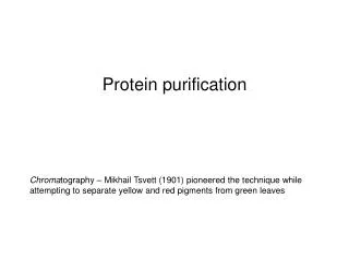
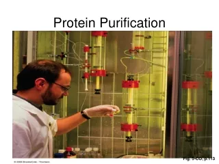
Chromatography • Chromatography: a broad range of physical methods used to separate and or to analyze complex mixtures. • The components to be separated are distributed between two phases: a stationary phase bed and a mobile phase which percolates through the stationary bed. paper chromatography stationary phase = paper mobile phase = solvent
Size-Exclusion Chromatography • Separation of proteins based on kinetics of moving through the available space (larger proteins have less space than smaller molecules) • Proteins larger than matrix elute in void volume (1 exchange of volume outside beads) • Proteins smaller than matrix partition in and out of beads • Pore size in beads is not uniform • Also some surface interaction with beads
Hydrophobic interaction chromatography • Hydrophobic group bound to solid phase • Binding • high salt (increases water surface tension, decreases available water molecules, increases hydrophobic interactions) • Elution • decrease salt • add detergent • decrease polarity of mobile phase
Affinity Chromatography • Ligand can be a small molecule, metal or antibody • Protein binds specifically to ligand attached to matrix • Elution with free ligand
Electrophoresis • Tris-glycine buffer • 10% SDS
coomassie brilliant blue A595 Electrophoresis • Protein detection • Coomassie blue • Sypro • Cybergreen • Silver staining
Using antibodies Antibodies (immunoglobulins) bind specific antigens/epitopes monoclonal - all bind same epitope polyclonal - mixture that binds several epitopes Secondary antibodies - anit-immunoglobulins (antibodies to antibodies)
Separate proteins by electrophoresis Transfer to membrane (e.g. nitrocellulose) Bind primary antibody Bind secondary antibody Detection Western blotting
Immuno-Affinity Chromatography • Antibody fixed to matrix • Protein binds to antibody • Wash unbound and loosely bound proteins off column • Elute protein with change in salt/pH
Protein purification simulation • http://www.tlsu.leeds.ac.uk/courses/bioc2060/proteinlab102/proteinlab.html
Example: Purification of Alkaline Phosphatase (AP) • Periplasmic Protein in E. coli • The space between the rigid peptidoglycan cell wall and the osmotically sensitive plasma membrane • Phosphate scavenger • Liberates Pi from a variety of substrates • Induced by phosphate starvation • Used to remove terminal phosphates for selective DNA ligation reactions • Heat stable, Zn enzyme
Assays • Enzymatic assays • PNPP is hydrolyzed to PNP and Pi • Fixed time assay • Mix enzyme and substrate, react for a fixed time, s • top the reaction with a strong base, • read the concentration of PNP at pH>10 • Continuous assay • Monitor PNP production directly in the spec at ph 8 • Bradford Assays for total protein • SDS page for the distribution of proteins by size.
Text Book Purification 1. Lysozyme treatment to release periplasmic proteins Centrifugation to separate soluble AP from cells Dialysis to remove starting buffer (overnight) 2. Heat treatment to precipitate weaker proteins Centrifugation to separate soluble AP from insoluble PPT Ammonium sulfate to concentrate proteins/remove non protein contaminants Dialysis to remove ammonium sulfate (O/N) 3. Anion exchange (DEAE) chromatography Step elution with 0.125M Salt 4. SDS Page to quantify the proteins in each fraction
Starting material E. coli cells starved for phosphate • Sucrose shrinks the plasma membrane reduces turgor pressure • Lysozyme cleave glycosidic linkages in cell wall • DNAse reduces viscosity from inadvertantly lysed cells • Left with AP, DNAse, Lysozyme, Sucrose other periplasmic and cytoplasmic contaminants
Alternative strategy Osmotic shock used to liberate periplasmic proteins • Many fewer proteins in periplasm than cytoplasm • Sucrose draws water from cytoplasm, shrinks inner membrane • EDTA permeabilizes cell wall • Transfer to low osmotic strength buffer causes the inner membrane to slam into the cell wall and force out periplasmic proteins • Periplasmic proteins, no lysozyme, no DNAase, not much sucrose