Download
1 / 1
10 likes | 92 Views
Jindrich Sedlacek 1, Jan Taraba2, Boris Cvek1 1 Department of Cell Biology & Genetics, Palacky University, Slechtitelu 11, Olomouc 78371 ; 2 Department of Chemistry, Episcopal High School, Barvicova 85, Brno 60200, Czech Republic. INTRODUCTION

E N D
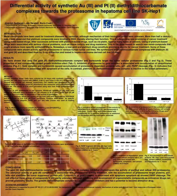
Jindrich Sedlacek1, Jan Taraba2, Boris Cvek1 1 Department of Cell Biology & Genetics, Palacky University, Slechtitelu 11, Olomouc 78371; 2 Department of Chemistry, Episcopal High School, Barvicova 85, Brno 60200, Czech Republic INTRODUCTION Metal compounds have been used for treatment diseases for centuries, although mechanism of their biological action were unknown. More than half a century ago, it was observed that platinum compounds were binding to DNA, therebyaltering their function. This is a beginning new promising of cancer treatment using metal-based drugs. After successes achieved with platinum-based drugs, such as cis-platin, against many types of cancers. Using of platinum-based drugs were associated with serious clinical problems, such as tissue toxicity and drug resistance. This is the main reason to search new metal-compunds that might produce more specific antitumor efects. Nowadays, a new gold and platinum drug constitute promising agents for cancer treatment.Some of these compoundswere shown activity against proteasome in various human tumor cell lines. We synthesized diethyldithiocarbamate complexes with platinum(II) and gold(III) and described them by X-raydiffraction and tested in hepato-carcinomaderived cell line SK-Hep1. RESULTS We have shown that only The gold(III)diethyldithiocarbamatecomplex anb bortezomib target the tumor cellular proteasome (Fig.-2 and Fig.-3). These properties of test compounds caused growth inhibition efect (Tab.-1). Inhibition of protesome by gold complex is associated with accumulation of ubiquitinated proteins (Fig.-5 ). Gold complex and bortezomib caused accumulation ofproteasome dependent proteins IκBα, p27, p53. PARP cleavage (Fig.- 6) indicates that proteasome inhibition is associated with apoptosis of the cells. In contrast, platinum (II) diethyldithiocarbamate complex is inactive in the tested parameters. Differential activity of synthetic Au(III) and Pt(II) diethyldithiocarbamate complexes towards the proteasome in hepatoma cell line SK-Hep1 METHODS Growth inhibition assay. Cells were cultured for 24 hours with synthetic coplexes. In parallel, the cells were treated with (DMSO; 0.001%) and Triton X-100 (5%) to assess the minimal and maximal cell damage, respectively. Cell viability was determined by MTT method using the standart protocol. Absorbance was measured spectrophotometrically at 570 nm. The values of IC50 were calculated using data from three independent cell passages. Inhibition of 20S proteasome in cell extracts. Whole-cell lyzates (10 μg) from cells treated as indicated were incubated for 120 minutes in 90 μl assay buffer with 20 μM substrate Suc-LLVY-AMC for detektion CT-like activity of proteasom. Immunoblot analysis. Whole-cell lyzates (50 μg) from cells were separated by SDS-PAGE and transferred to 0,45 0,45 μM PVDF membrane. Proteins were detekt by specific antibodies against ubiquitin, PARP, p53, p27and IκBα. β-actinwas used as loading control. Fig.-3. Inhibition of proteasomal CT-like activity in human SK-Hep1 cancer cells for 24 hours. Follows, cell extracts were prepared by lysis in buffer. Extracts (10 μg) were incubated with the fluorogenic substrate Fig.-2. Influence of 20S proteasome CT-like activity by [AuIII(DEDTC)3], [PtII(DEDTC)2], and bortezomib in cells lysat . Lysats (10 g) were preincubated with compounds for 15 minutes, followed by an additional 2-h incubation with the fluorogenic substrate Fig.-1.Molecular structures of both [AuIII(DEDTC)3] and [PtII(DEDTC)2] confirmed by using X-ray single-crystal diffraction Tab.-1. IC50 value of [AuIII(DEDTC)3], [PtII(DEDTC)2] and bortezomib were determined in SK-Hep1 cells after 24hours of treatment Fig.-6. PARP fragment (89 kDa). SK-hep1 cells were treated with complexes, bortezomib and solvent (DMSO)for 8 hours Fig.-5. Inhibition of degradation some important cellular proteinsIκB-α, p27, p53. Cells were treated withbortezomib, complexes and solvent (DMSO) for 8 hours Fig.-4. Accumulation of ubiquitinate proteins. SK- Hep1 cells were treated with solvent (DMSO), bortezomib and complexes for ½ hours CONCLUSION The gold (III) diethyldithiocarbamte complex, but not the platinum (II) complex, inhibited chymotrypsin-like activity of purified and celluar 20S proteasome. The antitumor activity of gold (III) complexes is associated with proteasomal activity inhibition, with the accumulation of proteasome target proteins, p27, IκBα and stabilizes the tumor suppressor protein p53. Cytotoxicity of gold complex is associated with apoptosis apoptosis as showed PARP cleavage. The cell effect of these group of compounds is still partly unknown, although screening and development of organometallic compounds as anticancer drugs can provide promising results. The gold(III) dithiocarbamate complex act as proteasome inhibitors and has a potential to become antitumor drug. ACKNOWLEDGMENT Our laboratory is supported by grant OP VK CZ.1.07/2.3.00/20.0062 “An inexpensive drug Antabuse as anticancer remedy: mechanism of action and clinical trials” from resources of European Union and the Czech Republic