Download
1 / 1
10 likes | 141 Views
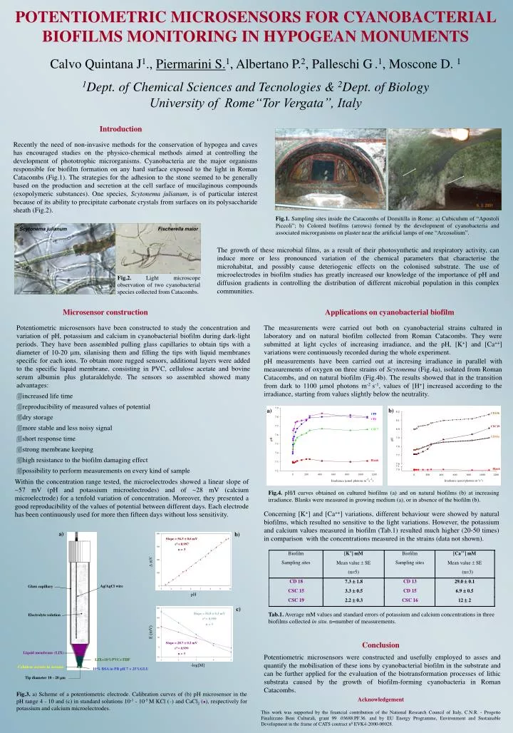
Fig.1. Sampling sites inside the Catacombs of Domitilla in Rome: a) Cubiculum of “Apostoli Piccoli”; b) Colored biofilms (arrows) formed by the development of cyanobacteria and associated microrganisms on plaster near the artificial lamps of one “Arcosolium”.

E N D
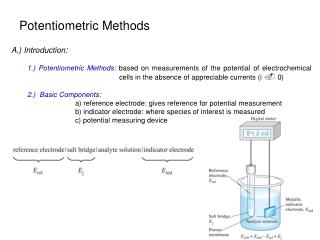
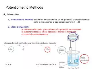
Fig.1. Sampling sites inside the Catacombs of Domitilla in Rome: a) Cubiculum of “Apostoli Piccoli”; b) Colored biofilms (arrows) formed by the development of cyanobacteria and associated microrganisms on plaster near the artificial lamps of one “Arcosolium”. Fig.2. Light microscope observation of two cyanobacterial species collected from Catacombs. Scytonema julianum Fischerella maior a) b) Slope = 56.5 ± 0.6 mV r2 = 0.997 n = 3 D mV Ag/AgCl wire Glass capillary pH c) Slope = 56.8 ± 0.3 mV r2 = 0.999 n = 3 Electrolyte solution a) b) E (mV) Slope = 28.7 ± 0.2 mV r2 = 0.999 n = 3 CD15b CP8 Liquid membrane (LIX) CP6 LIX+10%PVC+THF CSC19 -log[M] CSC7 Cellulose acetate in acetone 10% BSA in PB pH 7 + 25%GLU CD15a pH Tip diameter 10 - 20 m Blank Blank Irradiance (mmol photons m-2s-1) Fig.4. pH/I curves obtained on cultured biofilms (a) and on natural biofilms (b) at increasing irradiance. Blanks were measured in growing medium (a), or in absence of the biofilm (b). Tab.1. Average mM values and standard errors of potassium and calcium concentrations in three biofilms collected in situ. n=number of measurements. POTENTIOMETRIC MICROSENSORS FOR CYANOBACTERIAL BIOFILMS MONITORING IN HYPOGEAN MONUMENTS Calvo Quintana J1., Piermarini S.1, Albertano P.2, Palleschi G.1, Moscone D. 1 1Dept. ofChemical Sciences and Tecnologies & 2Dept. of Biology University of Rome“Tor Vergata”, Italy Introduction Recently the need of non-invasive methods for the conservation of hypogea and caves has encouraged studies on the physico-chemical methods aimed at controlling the development of phototrophic microrganisms. Cyanobacteria are the major organisms responsible for biofilm formation on any hard surface exposed to the light in Roman Catacombs (Fig.1). The strategies for the adhesion to the stone seemed to be generally based on the production and secretion at the cell surface of mucilaginous compounds (exopolymeric substances). One species, Scytonema julianum, is of particular interest because of its ability to precipitate carbonate crystals from surfaces on its polysaccharide sheath (Fig.2). The growth of these microbial films, as a result of their photosynthetic and respiratory activity, can induce more or less pronounced variation of the chemical parameters that characterise the microhabitat, and possibly cause deteriogenic effects on the colonised substrate. The use of microelectrodes in biofilm studies has greatly increased our knowledge of the importance of pH and diffusion gradients in controlling the distribution of different microbial population in this complex communities. Microsensor construction Applications on cyanobacterial biofilm Potentiometric microsensors have been constructed to study the concentration and variation of pH, potassium and calcium in cyanobacterial biofilm during dark-light periods. They have been assembled pulling glass capillaries to obtain tips with a diameter of 10-20 mm, silanising them and filling the tips with liquid membranes specific for each ions. To obtain more rugged sensors, additional layers were added to the specific liquid membrane, consisting in PVC, cellulose acetate and bovine serum albumin plus glutaraldehyde. The sensors so assembled showed many advantages: increased life time reproducibility of measured values of potential dry storage more stable and less noisy signal short response time strong membrane keeping high resistance to the biofilm damaging effect possibility to perform measurements on every kind of sample The measurements were carried out both on cyanobacterial strains cultured in laboratory and on natural biofilm collected from Roman Catacombs. They were submitted at light cycles of increasing irradiance, and the pH, [K+] and [Ca++] variations were continuously recorded during the whole experiment. pH measurements have been carried out at incresing irradiance in parallel with measurements of oxygen on three strains of Scytonema (Fig.4a), isolated from Roman Catacombs, and on natural biofilm (Fig.4b). The results showed that in the transition from dark to 1100 mmol photons m-2 s-1, values of [H+] increased according to the irradiance, starting from values slightly below the neutrality. Within the concentration range tested, the microelectrodes showed a linear slope of ~57 mV (pH and potassium microelectrodes) and of ~28 mV (calcium microelectrode) for a tenfold variation of concentration. Moreover, they presented a good reproducibility of the values of potential between different days. Each electrode has been continuously used for more then fifteen days without loss sensitivity. Concerning [K+] and [Ca++] variations, different behaviour were showed by natural biofilms, which resulted no sensitive to the light variations. However, the potassium and calcium values measured in biofilm (Tab.1) resulted much higher (20-50 times) in comparison with the concentrations measured in the strains (data not shown). Conclusion Potentiometric microsensors were constructed and usefully employed to asses and quantify the mobilisation of these ions by cyanobacterial biofilm in the substrate and can be further applied for the evaluation of the biotransformation processes of lithic substrata caused by the growth of biofilm-forming cyanobacteria in Roman Catacombs. Fig.3. a) Scheme of a potentiometric electrode. Calibration curves of (b) pH microsensor in the pH range 4 - 10 and (c) in standard solutions 10-1 - 10-5 M KCl () and CaCl2 (), respectively for potassium and calcium microelectrodes. Acknowledgement This work was supported by the financial contribution of the National Research Council of Italy, C.N.R. - Progetto Finalizzato Beni Culturali, grant 99 .03688.PF.36. and by EU Energy Programme, Environment and Sustainable Development in the frame of CATS contract n0 EVK4-2000-00028.