Download
1 / 87
870 likes | 906 Views
Orbit, accessory visual apparatus. Bony orbit

E N D
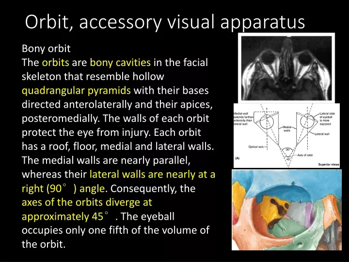
Orbit, accessory visual apparatus Bony orbit The orbits are bony cavities in the facial skeleton that resemble hollow quadrangular pyramids with their bases directed anterolaterally and their apices, posteromedially. The walls of each orbit protect the eye from injury. Each orbit has a roof, floor, medial and lateral walls. The medial walls are nearly parallel, whereas their lateral walls are nearly at a right (90°) angle. Consequently, the axes of the orbits diverge at approximately 45°. The eyeball occupies only one fifth of the volume of the orbit.
The remainder of the cavity is filled with vessels and verves that are comtained within and supported by orbital fat and connetive tissue. In brief, extraocular muscles; the optic, oculomotor, trochlear and abducent, and branches of the opththalmic and miaxillay divisions of the trigeminal nerve; the ciliary parasysmpathetic ganglion; the ophthalmic vessels; the nasolacrimal apparatus.
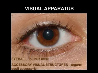
Orbit, Orbital Region, and Eyeball • The orbits contain and protect the eyeballs (globes of eyes) and accessory visual structures, which include the: • Eyelids • Extraocular muscles • Nerves and vessels • Orbital fascia • Mucous membrane (conjunctiva)
The pyramidal orbit has a base, four walls, and an apex The base of the orbit is outlined by the orbital margin that surrounds the orbital opening. The bone forming the orbital margin is reinforced to afford protection to the orbital contents and provides attachment for the orbital septum, an interrupted fibrous sheet that extends into the eyelids.
The pyramidal orbit has a base, four walls, and an apex The superior wall (roof) is approximately horizontal and is formed mainly by the obital plate of the frontal bone, which separates the orbital cavity from the anterior cranial fossa. Near the apex of the orbit, the superior wall is formed by the lesser wing of the sphenoid. Anterolaterally, a shallow depression in the orbital plate of the frontal bone, called the fossa for the lacrimal gland (lacrimal fossa), accommodates the lacrimal gland.
The pyramidal orbit has a base, four walls, and an apex Anteromedially it contains the frontal sinus and displays a small trochlear fovea, where the cartilaginous trochlea for superior oblique is attached. The optic canal lies between the roots of the lesser wing and bounded medially by the body of the sphenoid.
The pyramidal orbit has a base, four walls, and an apex The medial walls of the contralateral orbits are essentially parallel and are formed primarily by the orbital plate of the ethmoid bone, along with contributions from the frontal, lacrimal, and sphenoid. Anteriorly, the medial wall is indented by the lacrimal groove and fossa for the nasolacrimal sac. Much of the bone forming the medial wall is paper thin; the ethmoid bone is highly pneumatized with ethmoidal cells, often visible through the bone of a dried cranium.
The pyramidal orbit has a base, four walls, and an apex The ethmoid articulates with the medial edge of the orbital plate of the frontal bone at a suture which is interrupted by anterior and posterior ethmoidal foramina. Posteriorly, it articulates with the body of the sphenoid.
The pyramidal orbit has a base, four walls, and an apex The inferior wall (floor) is formed mainly by the orbital plate of the maxilla and partly by the zygomatic and palatine bones. The thin inferior wall is shared by the orbit and maxillary sinus. It slants inferiorly from the apex to the inferior orbital margin. The inferior wall is demarcated from the lateral wall of the orbit by the inferior orbital fissure, which connects the orbit posteriorly to the pterygopalatine fossa.
The pyramidal orbit has a base, four walls, and an apex The lateral wall is formed by the frontal process of the zygomatic bone and the greater wing of the sphenoid. This is the strongest and thickest wall, which is important because it is most exposed and vulnerable to direct trauma. Its posterior part separates the orbit from the temporal and middle cranial fossae. The lateral walls of the contralateral orbits are nearly perpendicular to each other. The lateral wall and roof are continuous anteriorly but are separated posteriorly by the superior orbital fissure.
The pyramidal orbit has a base, four walls, and an apex The apex of the orbit is at the optic canal in the lesser wing of the sphenoid just medial to the superior orbital fissure. The orbital canal connects orbit to the middle cranial fossa, transmitting the optic nerve and its meningeal sheaths, and the ophthalmic artery. The common tendinous ring, which gives origin to the four recti, is attached to the bone near the superior, medial and lower margins of the orbital opening of the canal.
Orbital connective tissue and fat Orbit contains a complex arrangement of connective tissue which forms a supporting framework for the eyeball and also acts to limit ocular rotation and compartmentalize orbital fat. This regions, including the orbital septum, fascial sheath of the eye, ‘check’ ligament, suspensory ligament and periosteum. * hyperthyroidism(Graves’ diseas)
Extraocular Muscles of the Orbit The extraocular muscles of the orbit are the levator palpelbrea superioris, four recti (superior, inferior, medial, and lateral), and two obliques (superior and inferior). These muscles work together to move the superior eyelids and eyeballs.. Muscles that are synergistic for one action may be antagonistic for another.
Levator Palpebrae Superioris The levator palpebrae superioris broadens into a wide bilaminar aponeurosis as it approaches its distal attachments. The superficial lamina attaches to the skin of the superior eyelid and the deep lamina to the superior tarsus. This muscle is opposed most of the time by gravity and is the antagonist of the superior half of the orbicularis oculi, the sphincter of the palpebral fissure. * Horner’s syndrome
Recti and Oblique Muscles The four recti muscles arise from a fibrous cuff, the common tendinous ring, that surrounds the optic canal and part of the superior orbital fissure. Structures that enter the orbit through this canal and the adjacent part of the fissure lie at first in the cone of recti. The lateral and medial recti lie in the same horizontal plane, and the superior and inferior recti lie in the same vertical plane.
Recti and Oblique Muscles From the primary position, the inferior oblique directs the pupil laterally and superiorly; therefore, when the superior rectus and inferior oblique work synergistically, pure elevation of the eyeball occurs. Similarly, the superior oblique directs the pupil inferiorly and laterally; therefore, when the superior oblique works synergistically with the inferior rectus, pure depression results.
Vascular supply and lymphatic brainage • The main vessel supplying orbital structures is the ophthalmic artery. Its terminal branches anastomose on he face and scalp with those of the facial, maxillary and superficial temporal arteries. The infraorbital branch of the maxillary arery, and the recurrent meningeal artery, also supply orbital structure. • Central retinal artey • Muscular branches • Ciliary arteries • Lacrimal artery • Supraorbital artery • Posterior and anterior ethmoidal artery; Meningeal branch; medial palpebral arteries; supratrochlear artery; dorsal nasal artery
veins • Superior ophthalmic veins • Inferior ophthalmic veins • Infraorbital vein • The superior and inferior ophthalmic veins link the facial and intracranial veins and are devoid of valves. • Cavernous sinus • Pterygoid venous plexus
Nerves of the Orbit The three terminal branches of the ophthalmic nerve, CN V1 (the frontal, nasociliary, and lacrimal nerves), pass through the superior orbital fissure and supply structures related to the anterior orbit (e.g., lacrimal gland and eyelids), face, and scalp. The cutaneous branches of CN V1 (lacrimal, frontal and infratrochlear nerves).
Nerves of the Orbit • The ciliary ganglion is a small group of postsynaptic parasympathetic nerve cell bodies associated with CN V1. The ganglion receives nerve fibers from three sources: • Sensory fibers from CN V1 via the communicating branch of the nasociliary nerve. • Presynaptic parasympathetic fibers from CN III • Postsynaptic sympathetic fibers from the internal carotid plexus.
Nerves of the Orbit The short ciliary nerves arise from the ciliary ganglion and are considered to be branches of CN V1. They carry parasympathetic (sphincter pupilae) and sympathetic fibers to the ciliary body and iris. The short ciliary nerves consist of postsynaptic parasympathetic fibers originating in the ciliary ganglion, afferent fibers from the nasociliary nerve that passthrough the ganglion, and postsynaptic sympathetic fibers that also pass through it.
Nerves of the Orbit Long ciliary nerves, branches of the nasociliary nerve (CN V1) that pass to the eyeball, bypassing the ciliary ganglion, convey postsynaptic sympathetic fibers to the dilator pupillae and afferent fibers from the iris and cornea.
Nerves of the Orbit In addition to the optic nerve (CN II), the nerves of the orbit include those that enter through the superior orbital fissure and supply the ocular muscles: oculomotor (CN III); trochlear (CN IV); and abducent (CN VI) nerves. A memory device for the innervation of the extraocular muscles moving the eyeball is similar to a chemical formula: LR6SO4AO3 (lateral rectus, CN VI; superior oblique, CN IV; all others, CN III).
Eyelids and Lacrimal Apparatus The eyelids are movable folds that are covered externally by thin skin and internally by transparent mucous membrane, the palpebral conjunctiva. This part of the conjunctiva is reflected onto the eyeball, where it is continuous with the bulbar conjunctiva. This part of the conjunctiva is thin and transparent and attaches loosely to the anterior surface of the eyeball. The bulbar conjunctiva, loose and wrinkled over the sclera (where it contains small, visible blood vessels), is adherent to the periphery of the cornea.
Eyelids and Lacrimal Apparatus • The upper eyelid is larger and more mobile than the lower eyelid, and contains an elevator muscle, levator palpebrae superioris. A transvers opening the palpebral fissure, lies between the free margins of the lids, which join at their extremities (medial and lateral canthus). • Lacrimal lake, triangular space • Lacrimal caruncle, reddish body • Plica semilunaris, a fold of conjunctiva • Lacrimal papilla • Punctum lacrimale • eyelashes
Eyelids and Lacrimal Apparatus Ectropion describes the rolling out of the lower eyelid so that it is no longer in contact with the cornea leading to epiphora. Entropion describes the inversion of the eyelid with corresponding inturning of the eyelashes (trichiasis) which contact the cornea and cause irritation. The anterior two thirds is skin and the posterior third is conjunctival mucosa.
Eyelids and Lacrimal Apparatus From its anterior surface inwards each eyelid consists of skin, subcutaneous connective tissue, fibres of the palpebral part of orbicularis oculi, submuscular connective tissure, the tarsal plate (tarsus) with its tarsal glands and orbital septum, and palpebral conjunctive. The subcutaneous connective tissue is very delicate, seldom contains any adipose tissue, and lacks elastic fibres.
Eyelids and Lacrimal Apparatus The superior (upper) and inferior (lower) eyelids are strengthened by dense bands of connective tissue, the superior and inferior tarsi (sing. tarsus) which form the "skeleton" of the eyelids. Embedded in the tarsi are tarsal glands, the lipid secretion of which lubricates the edges of the eyelids and prevents them from sticking together when they close. The lipid secretion also forms a barrier that lacrimal fluid does not cross when produced in normal amounts. When production is excessive, it spills over the barrier onto the cheeks as tears.
Eyelids and Lacrimal Apparatus • The eyelashes (L. cilia) are in the margins of the lids. The large sebaceous glands (of Zeis) associated with the eyelashes are Cilliary sweat gland (of Moll). The junctions of the superior and inferior eyelids make up the medial and lateral palpebral commissures, defining the angles of the eye.Thus each eye has medial and lateral angles, or canthi. • 睑腺炎(外麦粒肿),睑板腺炎(内麦粒肿),睑板腺囊肿(散粒肿)
Eyelids and Lacrimal Apparatus The lines of reflection of the palpebral conjunctiva onto the eyeball form deep recesses, the superior and inferior conjunctival fornices. The conjunctival sac is the space bound by the palpebral and bulbar conjunctivae; it is a closed space when the eyelids are closed, but opens via an anterior aperture, the palpebral fissure,when the eye is “open” (eyelids are parted). The conjunctival sac is a specialized form of mucosal “bursa” that enables the eyelids to move freely over the surface of the eyeball as they open and close. 沙眼 (trachoma)
Eyelids and Lacrimal Apparatus • The lacrimal apparatus consists of the: • Lacrimal glands: secrete lacrimal fluid (0.5-0.6ml/day, ). • Lacrimal ducts: convey lacrimal fluid from the lacrimal glands to the conjunctival sac. • Lacrimal canaliculi :commence at a lacrimal punctum (opening) on the lacrimal papilla near the medial angle of the eye and drain lacrimal fluid from the lacrimal lake (a triangular space at the medial angle of the eye where the tears collect) to the lacrimal sac (the dilated superior part of the nasolacrimal duct). • Nasolacrimal duct: conveys the lacrimal fluid to the inferior nasal meatus.
Eyeball The eyeball contains the optical apparatus of the visual system and occupies most of the anterior portion of the orbit. All anatomical structures within the eyeball have a circular or spherical arrangement. The eyeball proper has three layers; however, there is an additional loose connective tissue layer that surrounds the eyeball, allowing its movement within the orbit. The loose connective tissue layer is composed posteriorly of bulbar fascia, which forms the true socket for the eyeball, and anteriorly of bulbar conjunctiva.
Fibrous Layer of the Eyeball Fibrous layer (outer coat), consisting of the sclera and cornea. The sclera is the tough opaque part of the fibrous layer of the eyeball covering the posterior five sixths of the eyeball. The sclera accounts for approximately 93% of the outer coat of the eye. The anterior part of the sclera is visible through the transparent bulbar conjunctiva as "the white of the eye.“
Fibrous Layer of the Eyeball • Anterior scleral foramen • Limbus corneae • External and internal sulcus of sclera • Scleral spur • Sinus venosus sclera • Trabecular meshwork • Posterior scleral foramen • Cribriform plate of sclera
Fibrous Layer of the Eyeball • The cornea is the transparent part of the fibrous coat covering the anterior one sixth of the eyeball, it projects from the sclera as a dome-shaped elevation with an area of 1.1 cm2. The two parts differ primarily in terms of the regularity of the arrangement of the collagen fibers of which they are composed and the degree of hydration of each. • Microscopically, the cornea consist of five layers, corneal epithelium, anterior limiting lamina, substantia propia (stroma),posterior limiting lamina, and endothelium. • Tear film (lipid layer, aqueous phase, mucinous layer)
Fibrous Layer of the Eyeball • 角膜上皮不角化,角膜无上皮干细胞,上皮层更新依靠角膜缘的细胞向中心迁移。神经分部很丰富。 • 角膜云翳 (前界层形成瘢痕) • 角膜内皮,如损伤会导致角膜移植失败。
Vascular Layer of the Eyeball The vascular layer of the eyeball (also called the uvea or uveal tract) consists of the choroid, ciliary body, and iris. The choroid, a dark reddish brown layer between the sclera and the retina, forms the largest part of the vascular layer of the eyeball and lines most of the sclera. Within this pigmented and dense vascular bed, larger vessels of the vascular lamina are located externally. The finest vessels are innermost, adjacent to the avascular light-sensitive layer of the retina, which it supplies with oxygen and nutrients.
Vascular Layer of the Eyeball Engorged with blood in life, this layer is responsible for the “red eye” reflection that occurs in flash photography. The choroid is continuous anteriorly with the ciliary body. The choroid attaches firmly to the pigment layer of the retina, but it can easily be stripped from the sclera, and at the optic disc is continuous with the pia-arachnoid tissures around the optic nerve.
Vascular Layer of the Eyeball The ciliary body, which is muscular as well as vascular, connects the choroid with the circumference of the iris. The ciliary body provides attachment for the lens; contraction and relaxation of the smooth muscle of the ciliary body controls thickness (and therefore the focus) of the lens. Folds on the internal surface of the ciliary body, the ciliary processes, secrete aqueous humor, which fills the anterior and posterior chambers of the eye.
Vascular Layer of the Eyeball The ciliary body, which is muscular as well as vascular, connects the choroid with the circumference of the iris. The ciliary body provides attachment for the lens; contraction and relaxation of the smooth muscle of the ciliary body controls thickness (and therefore the focus) of the lens. Folds on the internal surface of the ciliary body, the ciliary processes, secrete aqueous humor, which fills the anterior and posterior chambers of the eye.
Vascular Layer of the Eyeball The iris, which literally lies on the anterior surface of the lens, is a thin contractile diaphragm with a central aperture, the pupil, for transmitting light. Two involuntary muscles control the size of the pupil: the parasympathetically stimulated sphincter pupillae closes the pupil, and the sympathetically stimulated dilator pupillae opens it. (2-8mm)
Vascular Layer of the Eyeball The iris, which literally lies on the anterior surface of the lens, is a thin contractile diaphragm with a central aperture, the pupil, for transmitting light. Two involuntary muscles control the size of the pupil: the parasympathetically stimulated sphincter pupillae closes the pupil, and the sympathetically stimulated dilator pupillae opens it. (2-8mm) The stroma of the iris is formed of fibroblasts, melanocytes; epithelial layers cell is a heavily pigmented cells
Vascular Layer of the Eyeball The anterior chamber of the eyeis the space between the cornea anteriorly and the iris/pupil posteriorly. The posterior chamber of the eye is between the iris/pupil anteriorly and the lens and ciliary body posteriorly.
Inner Layer of the Eyeball The inner layer of the eyeball is the retina. Grossly, the retina consists of two functional parts with distinct locations: an optic part and a non-visual retina. The optic part of the retina is sensitive to visual light rays and has two layers: a neural layer and pigment cell layer. The neural layer is light receptive. The pigment cell layer consists of a single layer of cells that reinforces the light-absorbing property of the choroid in reducing the scattering of light in the eyeball.
Cells of the retina Rod : rhodopsin(视紫红质) 498nm retinal(順式视黄醛)+opsin(视蛋白) (发色团,维生素A醛) 夜盲症nyctalopia 92 million Cone: iodopsin(视紫蓝质) 420nm blue, 534nm grenn, 563nm red
Inner Layer of the Eyeball The non-visual retinais an anterior continuation of the pigment cell layer and a layer of supporting cells over the ciliary body (ciliary part of the retina) and the posterior surface of the iris (iridial part of the retina), respectively.
Fundus of the eyeball The fundus is the posterior part of the eyeball. It has a circular depressed area called the optic disc (optic papilla) where the sensory fibers and vessels conveyed by the optic nerve enter the eyeball. Because it contains no photoreceptors, the optic disc is insensitive to light. Consequently, this part of the retina is commonly called the blind spot.