Download
1 / 27
280 likes | 554 Views
Muscles. Striated Cardiac Smooth Excitability and contractibility. animations. http://www.dnatube.com/video/4875/Physiology-of-muscle-contraction-and-relaxation http://www.dnatube.com/video/1306/Role-of-myosin-crossbridge-in-the-contraction-of-muscle

E N D
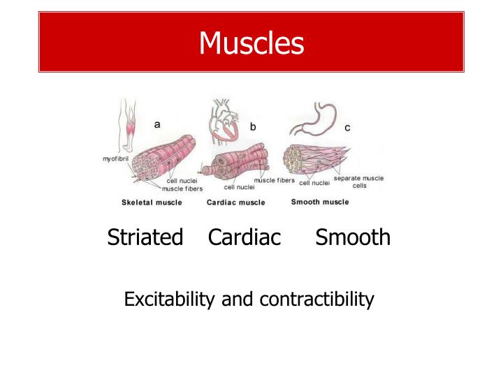
Muscles Striated Cardiac Smooth Excitability and contractibility
animations • http://www.dnatube.com/video/4875/Physiology-of-muscle-contraction-and-relaxation • http://www.dnatube.com/video/1306/Role-of-myosin-crossbridge-in-the-contraction-of-muscle • http://www.dnatube.com/video/1952/Sliding-filament-causes-contraction-of-muscle • http://www.dnatube.com/video/4154/Cellular-mechanism-of-muscular-contraction
Striated muscle – myography, tetanus • Muscle contraction • Twitch • Summation • Superposition • Tetanus • Smooth - multiple summation • Undulating – multiple superposition
Muscle strength • Muscle strength depends on the number of motor units recruited • Strength dependsonly on cross-sectional area20 – 100 N per sq.cmMuscle cells cannot divide. Thickening is formed by duplication of myofibrils. • Muscle strenght is influenced • genetically • hormonally – testosterone, anabolics
Muscle strength – tension/length curve, isometric and isotonic contraction
Sources of energy for muscle contraction • ATP – maintains contraction for 1 to 2 seconds • phosphocreatine – 5 times as great as ATP, sufficient for7-8 scontraction • Anaerobic Glycolysis • Enzymatic breakdown of the glucose to pyruvate and lactate liberates energy that is used to convert ADP to ATP, glycolysis can sustain contraction for about 1 min • Twofold importance of glycolysis • Reactions occurs in the absence of oxygen(muscle contraction can be sustained for a short time when oxygen is not available) • The rate of formation of ATP is2.5 times as rapid as ATP formation with oxygen • Oxidative metabolism– the final source of energy • 95% of all energy used by the muscle
Function of ATP ATP is necessary for • Muscle contraction – detachment of the head of myosin from the actin • Function of Na+/K+pump • Function of Ca++pump Physiological depletion of sources of ATP (reversible) – contracture, spasm, cramp Irreversible loss of all ATP – rigor mortis • Lack of energy for the separation of cross-bridges • Rigor is faster after muscle fatigue and exhaustion • Muscles remain in rigor until muscle proteins are destroyed by autolysis (15-25 hours)
Muscle fatigue • Acute (recovery - within 24 hours) and chronic (may be followed by a complete exhaustion) • Decrease force of muscle contraction • Fatigue • in the neuromuscular junction • Accumulation of extracellular K+ may lead to a disturbance in depolarization, reduction of the amplitude of the action potential and conduction velocity • decreasing amounts of muscle glycogen • Accumulation oflactate – lower pH, increase of K+, stimulation of the free nervous endings – pain, edemas • exhaustion of ATP
Muscle pain • After exercise • Dull ache when moving or being palpated • Begins in 1-3 days and lasts for one week • Maximal isometric strength is not impaired • Does not correlate with muscle edema, plasma CK, inflammation markers During exercise • Ischemic, hypoxic, accumulation of metabolites, pH • Fast in, fast out • Difficult to localize (muscle, bone, tendom, joint) • Referred pain (viscero somatic hyperalgesia)
Botulinum toxin preventsacetylcholine release– spasms (torticolis) Methacholine, carbachol and nicotine – the same effect as Ach – not destroyed by acetylcholinesterase – long action – Ophtalmology (glaucoma) Muscle relaxants– general anesthesia – muscle relaxation. Curare (D-tubocurarine) blocks acetylcholine receptorsw/o depolSuccinylcholine is a depolarizing blocker Anticholinesterase drugs, neostigmine and physostigmine– reversible inactivation of acetylcholinesterase – accumulaiton of Ach – myasthenia gravis Organophosphate – chemical weapons – irreversibleinactivation ofacetylcholinesterase– cramps, respiratory distress, sweating and convulsions. Dandrolen blocks Ca realease from SR – malignant hypetermia Drugs that modify neuromuscular junction
Smooth muscle - structure actin and myosin no troponin, calmodulin instead Dense bodies – analog of Z-lines – attachment of actin filaments Actin – long filaments, 15 times as myosin • Contraction 30 times slower than that of sceletal muscle • constant power during contraction (isotonic line longer,since some contractile units have optimal overlapping of A&M at one length of the muscle and others at other length)
Types of smooth muscles • Multiunite • discrete smooth muscle • single nerve ending • The ciliary muscle of the eye(parasympathetic control) • The piloerector muscles(sympathetic control) • Single-unit (visceral) • Hundreds to millions contract together– syncythial • gap junction – ions can flow freely • gut, bile ducts, ureters, uterus, vessels
Contraction of smooth muscle • Initiating event in smooth muscle contraction is an increase in intracelullar Ca2+ ions cause by: • Nerve stimulation • Stretch of the fiber • Hormonal stimulation • Changes in the chemical environment of the fiber • Strength of contraction depends on extracellular Ca2+ • Removal of Ca2+ ions is achieved by calcium pump, calcium pump is much slower in comparison with a pump of skeletal muscle – longer contraction
Mechanism of contraction • Beginning of contraction 4 Ca2+ bind with regulatory proteincalmodulin Complex Ca-calmodulin activatesenzyme myosin kinase (a phosphorylating enzyme) Light chain of of each myosin head (regulatory chain) become phosphorylated, the head has the capability of binding with the actin filaments • Cessation of contraction: When the concentration of Ca2+ falls bellow a critical level, all processes automatically reverse except for thephosphorylation of myosin head Enzymemyosin phosphatase splits the phosphate from the regulatory light chain
Smooth muscle – membrane potential • Resting potential –50 to –60 mV • Spontaneous slow wave (some smooth muscle is self-excitatory) • Slow wave can initiate action potentials (-35 mV) • The more AP, the stronger contraction Slow wave Smooth muscle has more voltage-gated calcium channels and very few voltage-gated sodium channels than skeletal m. Importance of Ca2+ ions in generating smooth muscle action potential – phase plateau of AP, contraction
Contraction without action potentials • In multiunite smooth muscle, Ca2+ions can flow into the cellthrough the ligand-gated Ca2+channel • ligand – acetylcholine, norepinephrine • Action potentials most often do not develop • Membrane potential do not reach a critical levelfor generating action potential because the Na+pump pumps sodium ions out of the cell
Regulation of smooth muscle Smooth muscle are regulated by autonomic nerves Nerve fibers do not make direct contact with smooth muscle fibers – they formed so-called diffuse junction Terminal axons have multiple varicosities, containing vesicules In the multiunite type of smooth cells, the contact junctions are similar to the end plate of skeletal muscle