Download
1 / 66
660 likes | 803 Views
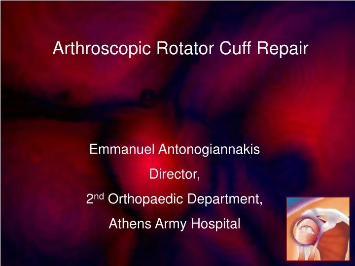
Arthroscopic Rotator Cuff Repair. Emmanuel Antonogiannakis Director, 2 nd Orthopaedic Department, Athens Army Hospital. First Description of RC tears.

E N D
Arthroscopic Rotator Cuff Repair Emmanuel Antonogiannakis Director, 2nd Orthopaedic Department, Athens Army Hospital
First Description of RC tears Smith JG. London. Med Gaz, 1834,14:280 Pathological appearances of seven cases of injury of the shoulder joint, with remarks. EA Codman Successful RC Repair Codman EA. Rupture of the supraspinatus tendon Boston Medical & Surgical Journal 1911 Vol clxiv (2) 708-10 McLaughlin HL. Lesions of the musculotendinous cuff of the shoulder: the exposure and repair of tears with retraction. J Bone Joint Surg 1944;26:31-51. HL McLaughlin
26 muscles controlling the shoulder girdle, only the four components of the rotator cuff are thought to play a significant role in the dynamic stability of shoulder joint The efficiency of the Concavity-Compression stabilizingmechanism 40% The most important stability mechanism in the mid-range motion, because the capsule and ligaments are lax in this range.
Rotator cuff disease is a spectrum of clinical conditions, which range from asymptomatic partial thickness tears to symptomatic rotator cuff arthropathy
The Gist of Operative Treatment • Stable RC repair • Restoration of tensile strength • Creation of an environment that facilitates healing mediated by the bursa • Prevention of bone/tendon gap formation Example of applied basic science in surgery
Supraspinatus Tendon • SS always involved • Five layer structure • Tear initiation in 85% • The size of the human supraspinatus tendon is 3.93 mm (France et al 1989) • Increased thickness and strength anteriorly (441-152-88 N) • Load to failure 650 N • Maximum generated force 200 N • Bursal side ε, strain, UTS < articular side • Different response- shearing effect(intratendinous tears)
RC Tendon Tear • Degenerated tissue (97% of tendon ruptures vs 34% control) • Chronic Tendinosis not Acute Tendinitis • Fibrocartilaginous metaplasia / Collagen type I ---III • Critical hypovascular zone distal 15 mm of SS-IS • Failure at the insertion • Poor stamina (Flag test, A.Castagna) Kannus, JozsaJ Bone Joint Surg, 1991,73,1507. Histopathological changes preceding spontaneous rupture of a tendon. A controlled study of 891 patients.
Partial tears More common than full thickness tears(13-37%) On the bursal side or the articular side of the rotator cuff. Usually get worse: Enlarge (52%) FTRCT (28%) Yamanaka, Matsumoto, CORR 1994
Why are articular side tears common ? Biological and Biomechanical explanation
Articular side: • vulnerable to tensile load • underperfused • UTS - strain to yield point 50% bursal side Υοung’s modulus, ε < < • Properties consistent cosistent proximal to distal • Different response to loading results in shearing effect between articular and bursal side Nakajima T. Histological and biomechanical characteristics of the supraspinatus tendon. J Shoulder Elbow Surg.,1994,3:79-87 Lohr, Uhthoff.The microvascular pattern of the supraspinatus tendon. Clin Orthop,1990,254:35-38
Bone trough (Mc Laughlin, 1944) • Proper environment for tendon healing • Increased distance for tendon insertion(greater tension) • Healing is not encouraged • Medialization of SS insertion • Internal impingement at the extremes of movement • Decortication decreases anchor pull out strength McLaughlin HLJ Bone Joint Surg 1944;26:31-51 Lesions of the musculotendinous cuff of the shoulder: the exposure and repair of tears with retraction
no significant benefit from the creation of a trough to expose the tendon to cancellous bone compared to cortical bone the biomechanical properties of the repair were significantly inferior than the intact control tendon Pierre et al. J Bone Joint Surg.77-A, 1995,1858-1866 Tendon-healing to cortical bone compared with healing to a cancellous trough. A biomechanical and histological evaluation in goats
The goal of the operative RC repair is to achieve a tension-free, water-tight repair. Is this possible ? Is this desirable ?
Calvert et al. J Bone Jt Surg 68B:147--150, 1986. Arthrography of the shoulder after operative repair of the torn rotator cuff. arthrography after operative repair of a torn rotator cuff in 20 patients(av.30 months pop) In 18 of 20 patients, contrast mediumleaked into the subacromial bursa indicating a defect in the cuff 17 patients no pain 15 full range of shoulder motion • No technique, however, has been immune from a recurrent tear • A watertight closure is not essential for a good functional result
Does debridement promotes healing of the tendon? Debridement may notstimulate the reparative process as was originally postulated, and their groupwere sceptical of the recovery of incompletely torn tendons to a load bearing functional structure after debridement of the cuff Kumagai J, Kawamata T, Sawai T. Rotator cuff disease- are-examination of the microanatomy and healing potential
Tendon ends are not avascular Power Doppler in RC tear Tear Debridement remove the ratty border of the tendon(1-2 mm)
How extensive debridement Bursa Resection Improved visualization + removal of inflammatory tissue but Might provide blood flow adding to the healing response Removal of superficial bursa only (Bigliani)
A chain with weak links Tendon Tendon to Suture Suture Suture to Anchor Anchor Anchor to Bone Bone Early failure is caused by suture or knot failure, suture pulling out of the tendon or through the bone.
Tendon to Suture Interface • No slippage - Cut off • Cyclic loading (gap formation - cycles to failure ) • multilooped suture techniques to grasp the tendon Mason-Allen suture technique locking technique locking loop technique
Which Suture • UTS • Stiffness • Elongation to failure • Knot formation • Slippage • Security of knots Braided polyester sutures(Ethibond-Tevdek) (Knots hold better ) Braided absorbable similar UTS but deterioration with time Monofilament sutures slip more easily and tying slip knots is easier
Suture to Anchor • Eyelet size • Low profile • Smooth edges • Suture Capacity • Unimpeded gliding
Anchor to Bone • Anchor design • Bone properties • Bone location • Usually load to failure > No 2 Ethibond • Worst case failure scenario: direction of pull is in the axis of insertion
Suture Anchor Pull-Out Strength • Tendon properties • Repair type • Suture type • Anchor • Bone Quality (osteoporosis-cortex-metaphysis-cancellous bone) • Mode of Loading(parallel to bone > perpendicular) • Metallic anchors more reliable
Bone • Elderly individuals • Osteoporotic bone (cheese wiring) • Reduced overall BMD of the involved extremity • Augmentation devices-Bone substitutes Deadman angle • Sulcus width > 2 mm - correlation with UTS • Clinical reports of anchors pulling out of the bone are noticeably lacking in the literature
But • The greater tuberosity showed anterior and posterior differences in anchor pullout force ( posterior 154 N, anterior 96 N) • Bone mineral density appears to have no correlation with the pullout strength of a screw-type suture anchor. FA Barber,SM Feder,SS Burkhart,J Ahrens. Arthroscopy 1999. The relationship of suture anchor failure and bone density to proximal humerus location: a cadaveric study
Which Anchor • Armamentarium • Clinical experience • Biomechanical competence • Biocompatibility • Long term in vivo behaviour • It is a matter of personal convenience
The ideal repair should have high initial fixation strength, allow minimal gap formation and maintain mechanical stability until solid healing France EP, Paulos LE, Harner CD, Straight CBAm J Sports Med 1989; 17:176-81. Biomechanical evaluation of rotator cuff fixation methods.
Rotator Cuff Repair Constructs • large single-pull loads suture failure • cyclic loading • biologicalfailure(bone failure or suture pullout from tendon)
Type of Loading and Mode of Failure RC - suture – anchor ---- Load to elongation Transosseous sutures suture breakage at the knot Cyclic Loading transosseous sutures vs suture anchors bone failure Anchors less prone to failure Burkhart et al. Arthroscopy, 1997,13,720 Cyclic loading of anchor-based rotator cuff repairs: confirmation of the tension overload phenomenon and comparison of suture anchor fixation with transosseous fixation.
Strength of Repair Bone - Tendon complex 6 wks 30% 3 months 52% 6 months 81% Failure to protect the RC repair results in high failure rates Low load cyclical loading causes gap formation Gerber et al. J Bone Joint Surg. 1999,81,1281 Experimental rotator cuff repair. A preliminary study.
Six methods of fixation to bone Gerber et al. J Bone Joint Surg. 76-B, No. 3,1994, 371-380 Mechanical strength of repairs of the rotator cuff.
Cortical graft 140 Tendon pulled out Metallic brush 299 Tendon pulled out Double transosseous fixation 146 Suture pulled out Single Transosseous fixation 139 Suture pulled out Mitek G II anchor 142 Anchor pulled out Membrane augmentation 329 Suture ruptured Gerber et al. J Bone Joint Surg. 76-B, No. 3,1994, 371-380 Mechanical strength of repairs of the rotator cuff.
Transosseous sutures cut through bone at 150 N PL plate augmentation at 329 N Cyclic loading data show that sutures in bone tunnels can fail when subjected to as few as 25 cyclic loads at physiologic levels Burkhart SS, Johnson TC, Wirth MA, Athanasiou KA Arthroscopy 1997;13:172-176. Cyclic loading of transosseous rotator cuff repairs: Tension overload as a possible cause of failure.
Mod Mason-Allen 359 Mod 1, Kessler 329 Kleinert 338 Mod 2, Kessler 350 Mattress 269 Locking 392 Mod 3, Kessler 366 Kleinert augmented 329 Locking loop 383 Simple stitch (6 stitches) 273 Mod Mason-Allen augmented 389 Mod 1, Kessler augmented 229 Simple stitch (4 stitches) 208 Simple stitch (2 stitches) 184 Ultimate tensile strength(N)
Most techniques, other than the simple stitches, caused definite deformation of the tendon by the loaded and tightened sutures. Modified Kessler Mason-Allen
Mason-Allen grasping suture Only the modified Mason-Allen technique withstood 1000 cycles There was no slippage Progressive longitudinal pulling caused increased transverse tightening of the suture loops
The augmentation of the techniques with an absorbable poly(L-/D-lactide) membrane did not improve holding strength. The membrane acted as a gliding plane and did not allow the suture to grasp the fibre bundles of the tendon; the results were weaker and showed larger separations.
Simple Stitch • Is surprisingly weak • Does not strangulate the tendon • Allows almost no separation in the absence of significant tension • For small cuff tears in which reinsertion to bone can be obtained without tension and the repair can be protected from tensile load during early healing • Not adequate for larger tears
Elongation under Ultimate tensile strength (N) 40 N load (mm) Ethibond 3 6.5 106 Mersilene 2 7.5 83 Ethibond 2 7.9 82 Vicryl 2 10.5 90 PDS II 2 20.6 109 Ethibond 1 9.8 65 Vicryl 1 11.6 66 PDS II 1 28.3 85 Ethibond 0 10.9 54 Vicryl 0 16.8 52 Dexon Plus 0 17.0 53 PDS II 0 37.4 59 Prolene 0 45.6 42 Ethilon 0 48.4 41
For the same suture material, one size larger increased the UTS from size 0 to 1 35% 1 to 2 30%.
Reed et al. Am J Sports Medicine1996 Full-Thickness Rotator Cuff Tears. A Biomechanical Comparison of Suture Versus Bone Anchor
The repairs were loaded cyclically and failed at low loads by cutting into bone and tendon, casting doubt on the integrity of the repair in early mobilization after surgery.
Load-versus-displacement graph for cyclic loading (suture anchors in lateral cortex, 45 degrees to surface) Permanent incremental deformation with each load cycle and a larger elongation during the step-up to a larger maximum force Elongation 7 mm between the first and the 20th load cycles. Elongation 20 mm between the first and final loadings
Typical appearances of repairs after cyclic loading (a)with and (b) without suture anchors. The sutures have sawn into the bones and torn into the tendon. The suture anchors did not move.
Higher load-to-failure strengths may be of questionable importance because the limiting factor is still the suture strength. • Choosing an anchor with a pullout strength of 125 lb over another anchor with a pullout strength of 60 lb solely because of the load to failure data ignores the fact that a No. 2 suture placed in either anchor will break by the time the load on it reaches 30 lb; • In all likelihood, before the suture broke, it will have torn through the soft tissue first F.A.Alan Barber, Morley A. Herbert. Arthroscopy, 15,1999. Suture Anchors-Update 1999
Anchors resumeé • Screw anchors have higher values, but for the newer nonscrew designs this distinction is less apparent • The new biodegradable anchors are all composed of poly L- lactic acid • All anchors were stronger than the suture for which they are designed to accommodate F.A.Alan Barber, Morley A. Herbert. Arthroscopy, 15,1999. Suture Anchors-Update 1999
Unstable fulcrum(SS-IS) Stable fulcrum(SS-part IS)