Download
1 / 1
50 likes | 637 Views
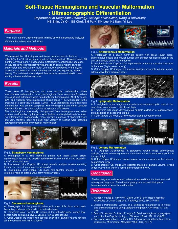
Soft-Tissue Hemangioma and Vascular Malformation : Ultrasonographic Differentiation Department of Diagnostic Radiology, College of Medicine, Dong-A University HS Shin, JY Oh, SS Choi, BH Park, KN Lee, KJ Nam, YI Lee. Purpose.

E N D
Soft-Tissue Hemangioma and Vascular Malformation: Ultrasonographic DifferentiationDepartment of Diagnostic Radiology, College of Medicine, Dong-A UniversityHS Shin, JY Oh, SS Choi, BH Park, KN Lee, KJ Nam, YI Lee Purpose To differentiate the Ultrasonographic findings of Hemangioma and Vascular Malformation arising from soft tissue C B A Materials and Methods Fig.3.Arteriovenous Malformation A. Photograph of a seven month-old patient with about 4x2cm sized, erythematous nodule and irrgular surface with purplish red discoloration of the skin and located below the left auricle. B. Longitudinal color Doppler US image reveals numerous vascular structures in the mass.( high vessel density) C. Color Doppler US image with spectral analysis of sample volume reveals arterial wave form within a vessel. We reviewed the US findings of soft tissue vascular mass in thirty six patients( M:F = 19:17) ranging in age from three months to 13 years (mean 36 months). Among them, 14 cases were histologically confirmed by operation. The 22 cases were retrospectively diagnosed by clinical history, physical examination and involution of lesion during follow-up period. We analyzed presence of solid tissue, abnormal artery and vein, echogenecity, vessel density. The resistive index and peak flow velocity were evaluated in mass, feeding arteries and draining veins. Results There were 27 hemangioma and nine vascular malformation (three arteriovenous malformation, three lymphangioma, three venous malformation). The significant differences were noted between hemangioma (24 of 27 cases, 88%) and vascular malformation (one of nine cases, 11%) with respect to the presence of a solid tissue mass(p< .001). The vessel density of arteriovenous malformation was greater compared with hemangioma and other vascular malformation such as lymphangioma or venous malformation. The lymphangioma was easily distinguished from hemangioma and other vascular malformation by the poor vascularities, multiseptated cysts in mass. No differences in echogenecity, vessel density, presence of abnormal artery and vein, resistive index and peak flow velocity of vessles were detected between hemangioma and vascular malformation. B A C Fig.4.Lymphatic Malformation A. T1 weighted coronal image demonstrates multi-septated cystic mass in the subcutaneous portion of buttock and thigh. B. Transverse US image demonstrates multiple collection of subcutaneous cysts septated by echogenic stroma. C. Color Dopple US reveals a few vesseles along echogenic septa. A B C C A B Fig.5.Venous Malformation A. T1 weighted Gd-enhanced fat suppresed coronal image demonstrates multiple, tortous enhancing vascular structures in the subcutaneous portion of the right thigh. B. Color Doppler US image reveals several venous structure in the mass on compression view. C. Color Doppler US image with spectral analysis of sample volume reveals venous wave form within a vessel on compression view. Fig.1. Strawberry Hemangioma A. Photograph of a nine month-old patient with about 2x2cm sized, erythematous nodule and purplish red discoloration of the skin and located in the left infraorbital area. B. Transverse color Doppler US image reveals multiple vessles coursing through the mass ( moderate vessel density) C. Longitudunal color Doppler US image with spectral analysis of sample volume reveals an arterial wave form within a vessel. Conclusion The hemangioma and vascular malformation are different in treatment and subsequent prognosis. The ultrasonography can be used distinguish hemangioma from vascular malformation. Reference B C A 1. Harriet J, Patrica E, Harry P.W, David, John B. Soft-Tissue Vascular Anomalies of US for Diagnosis. Radiology 2000; 214:747-754 2. Dubois J, Patriquin HB, Garel L, et al. Softtissue hemangiom as in infants and children: diagnosis using Doppler sonography. AJR 1998; 171:247-252 3. Bulas DI, Johnson D, Allen JF, Kapur S. Fetal hemangioma: sonographic and color flow Doppler findings. J Ultrasound Med 1992; 11:499-501. 4. Cohen JM, Weinreb JC, Redman HC. Arteriovenous malformations of the extremities: MR imaging. Radiology 1986; 158:475-479 Fig.2.Cavernous Hemangioma A. Photograph of a five year-old patient with about 1.2x1.2cm sized, soft-fluctuating nodule and located in the left-sided nose. B. Transverse color Doppler US image of the left-sided nose reveals low-echoic mass containing several vessles.( low vessel density) C. Color Doppler US image with spectral analysis of sample volume reveals an arterial wave form within a vessel.