Download
1 / 53
530 likes | 565 Views
Conduct of anesthesia. The physiologic state induced by general anesthetics typically include analgesia , amnesia , loss of consciousness , hypnosis , inhibition of sensory and autonomic reflexes and skeletal muscle relaxation. ( Guedel’s signs ) and stages of anesthesia

E N D
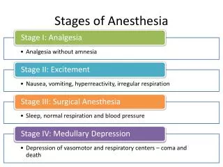
The physiologic state induced by general anesthetics typically include analgesia , amnesia , loss of consciousness , hypnosis , inhibition of sensory and autonomic reflexes and skeletal muscle relaxation. • ( Guedel’s signs ) and stages of anesthesia • 1- Stage of Analgesia ( induction ) and amnesia. • 2- Stage of Excitement ( Delirium ) and loss of consciousness. • 3- Stage of surgical anesthesia. • 4- Stage of medullary depression and failure of circulation (death)
Stage 1 :- From beginning of induction to loss of consciousness. Stage 2 :- From loss of consciousness to onset of automatic breathing. Eyelash reflex disappear , Coughing, vomiting may occur , Respiration can be irregular with breath-holding. Stage 3 :- READY FOR SURGERY From onset of automatic respiration to respiratory paralysis. Plane I - from onset of automatic respiration to cessation of eyeball movements. Plane II- from cessation of eyeball movements to beginning of paralysis of intercostal muscles. secretion of tears increases (a useful sign of light anesthesia). Plane III- from beginning to completion of intercostal muscle paralysis Plane IV- from complete intercostal paralysis to diaphragmatic paralysis . Stage 4 :- from stoppage of respirationtill death Pupils dilated, muscles relaxed.
Phases of anesthesia • A- Induction • B- Maintenance • C- Recovery
Induction :- The period from onset of administration of the anesthetic to development of effective surgical anesthesia. It depends on how fast the anesthetic drug reaches the brain. • Indications • No intravenous access • (neonates ,infants, children) • Perioperative anxiety or needle phobia • Procedural sedation ( nitric oxide ) ( emergency , dentistry , endoscopy ) • Maintenance of anesthesia after administration of an intravenous agent. • Cesarean section and delivery • Air way obstruction ( epiglottitis ) • Bronchopleural fistula or empyema.
1- Rapid induction and emergence ( low solubility ). ------ reversible 2- Allow the concomitant administration of high oxygen. 3- Painless. 4- Enhanced safety and recovery. 5- Doesn’t need IV access. 6- Easy to administer. 7- The depth of anesthesia can be rapidly altered by changing the concentration of the drug Advantages of inhalational induction
Difficulties and complications • 1- Laryngeal spasm • Sustained closure of the vocal cords resulting in the partial or complete loss of the patient's airway by constriction of the intrinsic laryngeal muscles • Airway reflex that protects against aspiration but can occur in light planes of anaesthesia • Through Direct laryngeal or distant visceral stimulation • Leads to hypoxaemia and bradycardia • Prevention, recognition , increase depth of anesthesia , post anesthetic care. • Due to :- • Airway irritation Tonsillectomy and adenoidectomy • Volatile anaesthetics Thyroid surgery • Manipulation ( Laryngoscopy, suction catheter ) Smoking • Inexperienced anesthetists URTI • young children at greatest risk Asthma
2- Airway obstruction (apply continuous positive airway pressure ) • Upper : falling back of tongue , foreign body , endobronchial intubation , hiccups • 3- Bronchospasm • Causes :- allergy, smokers, irritants , URTI • treatment :- increase depth of anesthesia with additional induction agent or volatile agent, IV or endotracheal lidocaine, humidification and warming of gases,bronchodilators. • 4- Slow induction of anesthesia • 5- Environmental pollution ( Scavenger system is needed to collect exhaled anesthesia , medications or gases ) 6- Seizures during induction ( enflurane)
7- Malignant Hyperthermia • Hyperpyrexia • Genetic • Life threatening • Hypermetabolic disorder of skeletal muscle • Triggered by general anesthetics and succinylcholine • Treatment : Dantrolene
Signs and symptoms of malignant hyperthermia • Non specific • Tachycardia • Tachypnea • Arrythmia • Hypotension • Hypertension • Cyanosis • Metabloic acidosis • Hyperkalemia • Specific • Muscle rigidity • Fever ( late ) • Myoglobinemia ( late ) • Increase expired Co2 • Increase serum creatine phosphate ( late )
Procedure S : Suction checked and functioning. A : Airway equipment checked and prepared. (functioning and backup laryngoscope, ETT , oxygen source and manual resuscitation bag). M :anaesthesia Machine M : Monitors M : Medications • Anesthesia can be induced by having the patient breathe increasing concentrations of inhaled gases by mask.
1- The mask is introduced gradually to the face from the side. 2- Pull the chin up 3- Position the patient 4- Preoxygenationwith 100% oxygen for 3-4 minutes lessen the risk of hypoxemia occurring during the apneic period after induction 5- Anesthetist adjusts the mixture of gas flow &observes the patient’s reaction. initially No2 70% in O2 ( carrier gas ) is used. 6-Anesthesia deepened by gradual introduction of a volatile agent. - A single breath technique for patientswho cooperate. ( inc.vapour concentration) 7- Observe the pattern of ventilation, palpate peripheral pulses, monitor. 8- Insertion of a laryngeal mask airway or tracheal tube may be considered when anesthesia has been established.
Maintenance : • the period between the start of the surgical intervention up to when it was complete. • If no further agents were administered following the induction of anesthesia the patient would awaken within minutes. Therefore, maintenance of anesthesia requires the delivery of pharmacologic agents with the aim of achieving the “four A’s of anesthesia” and hemodynamic stability throughout the surgical procedure.
Maintenance of general anesthesia is typically achieved by: inhaled anesthetics (Sevoflurane, Isoflurane but also Halothane 1-2 MAC may be employed in a mixture of nitrous oxide 70% in oxygen). intravenous drugs (TIVA:Propofol and Opioid) IV opioids (Fentanyl, Alfentanil, Remifentanil)
Depends on : • Nature and length of surgery. • Provision of analgesia in the premedication. • Patient’s response (ventilation ,circulation, HR & rhythm). • Tracheal intubation.
Conduct of inhalational anesthesia with spontaneous ventilation. Appropriate form of maintenance for: 1. Superficial operations. 2. Minor procedures which produce little reflex or painful stimulation. 3. Operations for which profound muscle relaxation is not required. Difficulties and Complications:1. Airway obstruction 2. Laryngeal spasm 3. Bronchospasm 4. Malignant hyperthermia: 5.Raised intracranial pressure (Inhalational > IV). 6. Awareness
Awareness complication of anesthesia whereby a patient who has received a general anesthetic becomes conscious of his or her environment during the surgical procedure. The patient may or may not experience pain and may or may not recall the events post-operatively. • It may be prudent to warn such patients of the risk pre-operatively in high risk. Risk factors: Light anesthesia lifestyle factors like drinking or abusing drugs, and medications being taken by the patient. Emergency surgery. Difficult airways. Machine malfunction or misuse Human errors.
The risk of awareness is reduced by avoidance of paralytics unless necessary; careful checking of drugs, doses and equipment; good monitoring, and careful vigilance during the case.
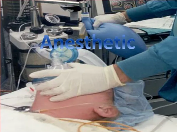
Airway maintenance delivery of inhalation agents: 1. Face Mask: • Applied before and during and after loss of consciousness at anesthetic induction. The mandible is held into the mask by the anesthetics (holding rather than pressing)-the mandible is held foreword , helping to prevent posterior movement of the tongue and obstruction of the airway. •It has variants of type and size.
Airway obstruction may occur in variety ways:The most common cause in an unconscious supine :patient is from the *tongue falling back into the hypopharynx*decrease in the tone of muscles attaching the tongue to the mandible, hyoid bone and epiglottis*The respiratory efforts of the unconscious patient tend to pull the tongue backward causing further airway obstruction.
Excluding intubation, simple maneuvers to overcome upper airway obstruction in the unconscious supine patient, include:1. Clearing the airway of any foreign material2. Using a chin lift maneuver.(lift the patient’s tongue away from the back of the throat and provide an adequate airway)3. Using a jaw thrust maneuver. 4. Inserting artificial airways: oropharyngeal, nasopharyngeal or laryngeal mask airway.5. Positioning the patient on their side in the semi-prone recovery position
Jaw Thrust Maneuver Chin Lift Maneuver
Oropharyngeal airway (guedel)• Curved plastic tubes, flattened in cross-section and flanged at the oral end. They lie over the tongue, preventing it from falling back into thepharynx.
• An estimate of the size required is given by comparing the airway length with the vertical distance between the patient’s incisor teeth and the angle of the jaw.
• Available in a variety of sizes suitable for all patients,from neonates to large adults. The commonest sizes are 2–4, for small to large adults, respectively• Initially inserted ‘upside down’ as far as the back of the hard palate rotated 180° . and fully inserted until the flange lies in front of the teeth, or gums. .
**The device is removed when the person regains gag reflex and can protect their own airway. **Simply remove by pulling on it withoutrotation **Can facilitate ventilation during CPR (cardiopulmonary resuscitation) and for persons with a large tongue
Nasopharyngeal airway **also known as nasal trumpet (because of itsflared end), or nose hose ** Round, malleableplastic tubes, The purpose of the flared end is to prevent the device from becoming lost inside the patient's nose
**A guide to the correct size is made by comparing the diameter to the external nares. The diameter of the airway should be the largest that will fit The common sizes in adults are 6–8 mm, for ** Small to large adults, Respectively **Prior to insertion, the patency of the nostril (usually the right) should be checked
NOTE: Nasal airways are contraindicated in-patients ** with severe trauma to the head and/or face due to the possibility of direct intrusion into brain tissue ** If an obstruction is encountered, do not force the airway as severe bleeding may be provoked. The airway should be removed and .inserted in the left nostril
2. The Laryngeal Mask Airway: The laryngeal mask airway is a specialized airway device made of wide bore PVC tubing, which incorporates a distal inflatable laryngeal cuff. The LMA is inserted without special equipment, in the back of the patient's pharynx with the soft laryngeal cuff resting above the vocal cords at the junction of the larynx and esophagus.
Indications: a. Provide clear airway without the need to support a mask. b. Avoid the use of tracheal intubation during spontaneous ventilation. c. In a case of difficult intubation , to facilitate subsequent insertion of a tracheal tube. • Contraindications: a. Full stomach or any condition lead to delayed gastric emptying. b. Possible regurgitation . c. Surgical access is Impeded By the cuff of the LMA . d. Thoracic surgery. e. an unusual position (Prone). • f. Patients with oropharyngeal or retropharyngeal pathology, or foreign bodies in the hypopharynx.
Technique for insertion of thestandard LMA • The patient’s reflexes must be suppressed to a level similar to that required for the insertion of an oropharyngeal airway to prevent coughing or laryngospasm • The cuff is deflated and the mask lightly lubricated • A head tilt is performed, the patient’s mouth opened fully and the tip of the mask inserted along the hard palate with the open side facing but not touching the tongue • The mask is further inserted, using the index finger to provide support for the tube. Eventually, resistance will be felt at the point where the tip of the mask lies at the upper oesophageal sphincter • The cuff is now fully inflated using an air-filled syringe attached to the valve at the end of the pilot tube • The laryngeal mask is secured either by a length of bandage or adhesive strapping attached to the protruding tube
TRACHEAL INTUBATION • the placement of a flexible plastic tube into the trachea to maintain an open airway or to serve as a pathway for drug administration. • tracheal intubation is employed both for the conduct of general anesthesia and to facilitate the ventilator management of the critically ill. • Intubation is not a risk-free procedure, and it is not a requirement for all patients receiving general anesthesia .
Indications for Intubation 1- Inability to protect the airway against aspiration ( in order to Protect the respiratory tract from aspiration of gastric contents in patients who are at risk ) . 2-those undergoing surgical procedures involving body cavities ( head and neck ) . 3- Surgical procedures within the chest, abdomen . 4- those who will be positioned so that the airway will be less accessible (eg , those undergoing surgery in the prone position, or whose head is rotated away from the anesthesia workstation ).
5- Inability to maintain airway patency. 7- Failure to ventilate. 8- Failure to oxygenate. 9- Anticipation of a deteriorating course that will eventually lead to respiratory failure.
Preparation • 1-checking equipment and properly positioning the patient. • 2-checking the patency of the tracheal tube . • 3-laryngoscope handle, and bulb function should be tested . • 4- should have a dedicated and experienced assistant .
A rigid laryngoscope The sniffing position and intubation
Steps for Tracheal Intubation * Step1 : Check the equipment and Assemble all materials close at hand . * Step2 : Position of the patient - Elevating the patient’s head about 10 cm with pads under the occiput and extension of the head into the sniffing position - (This position permits better visualization of the glottis and vocal cords and allows easier passage of the endotracheal tube ) . * Step3 : Curved blade technique . • A . Hyper-oxygenate the patient with 100% oxygen for 2 minutes. • B . Open the patient’s mouth with the right hand, Grasp the laryngoscope in the left hand. Spread the patient’s lips, and insert the blade between the teeth, being careful not to break a tooth. • C . Pass the blade to the right of the tongue, pushing the tongue to the left. • D . Lift the laryngoscope upward and forward, without changing the angle of the blade, to expose the vocal cords . apply gentle downward pressure on the cricoid cartilage, start off slowly and then gradually increase the downward force. • E . Pass the tube through the vocal cords.
Steps for Tracheal Intubation cont. • F . Inflate the tube Cuff with proper volume ( adequate to provide the seal, and not exceeding not exceeding 30 cm HO2 ) • G . Ventilate through the tube and check that both lungs are equally ventilated by auscultating both lungs . • H . The tube distal end should ideally be 1 to 2 cm above the Carina , otherwise right bronchial intubation is likely with only the right lung being ventilated . • I . Firmly secure the tube by plaster taping it to the face or by the use proper tape .
An endotracheal tube in good position on CXR. Arrow marks the tip An endotracheal tube not deep enough. Arrow marks the tip
Anesthesia for tracheal intubation • It can be done: • A . Under general anesthesia (either i.v. or inhalational +\-muscle relaxation). • B . Awake Intubation under local anaesthesia using (topical spry , transtracheal spry and superior laryngeal nerve block).
Anesthesia for tracheal intubation cont. • Inhalational technique for intubation:- • Adequate depth of anaesthesia is necessary to depress the laryngeal reflexes and to provide muscles relaxation. • Using halothane in concentrations up to 4% may provide the necessary depth. • The adequate depth can be confirmed when there is predominance of diaphragmatic breathing(the chest does not rise and the belly expands during this type of breathing ) .
Anesthesia for tracheal intubation cont. • Relaxant Anesthesia for intubation: •After i.v. or inhalational induction of anesthesia , muscle relxant (example: suxamethonium short acting depolarizing agent) may be used to provide muscle relaxation for intubation . Suxamethonium is administered in a dose of 1 to 1.5mg/kg. •Assisted ventilation is maintained via the face mask until muscle relaxation occurs ( except in emergency patients and those likely to regurgitate). •Then the mask is removed and laryngoscopy and intubation performed . The anesthetic circuit is then connected to the tracheal tube and anesthetic maintained at a depth appropriate for surgery .
Anesthesia for tracheal intubation cont. • Indications of Relaxant Anesthesia for intubation It provides muscle relaxation, permitting anesthesia for: 1-major abdominal, intra peritoneal, thoracic or intracranial Operations . 2-prolonged operations . 3-prolonged ventilation .
Complications of intubation 1- malpositioning . 2- airway trauma . - dental damage , sore throat . - lip , tongue , mucosal laceration . - dislocated jaw . - retropharyngeal dissection . 3- physiological reflexes . - hypoxia , hypercarbia . - hypertension , tachycardia . - intraocular and intracranial hypertension . - laryngospasm . 4- tube malfunction ( cuff perforation ) .
EXTUBATION • Extubation refers to removal of the endotracheal tube (ETT). It is the final step in liberating a patient from mechanical ventilation . • Tracheal extubation is a high risk procedure in anaesthesia and critical care. • Extubation should not be performed until it has been determined that the patient’s medical condition is stable. • extubationshould be performed when a patient is either deeply anesthetized or awake , Extubation during a light plane of anesthesia (ie, a state between deep and awake) is avoided because of an increased risk of laryngospasm .