Download
1 / 31
310 likes | 318 Views
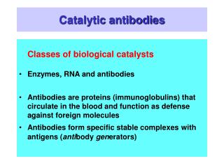
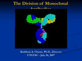
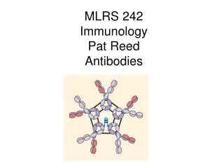
Monoclonal Antibodies (mAbs) Antibodies (Abs). Also known as immunoglobulins (Ig). Comprised of 2 heavy chains and 2 light chains Monoclonal Abs bind specifically to a single site (epitope) on a particular antigen Abs are produced by B lymphocytes.

E N D
Monoclonal Antibodies (mAbs) • Antibodies (Abs). Also known as immunoglobulins (Ig). • Comprised of 2 heavy chains and 2 light chains • Monoclonal Abs bind specifically to a single site (epitope) on a particular antigen • Abs are produced by B lymphocytes. • Because of their specificity and ease of generation, they are extensively used as therapeutics (“passive immunotherapy”) and as diagnostic and research tools • - They can be generated in large (unlimited) amounts in culture
Antibodies are made by B-cells B cells develop in the bone marrowhematopoietic stem cells and lymphoid stem cells lymphoid stem cells T-cells and B-cells progenitor = pro-B cell (B220+) precursor = pre-B cells: heavy-chain rearranged immature B cell: IgM + light-chain rearranged matured B cell: IgM + IgD + an antigen encountered in spleen or lymph nodes; then goes to peripheral circulation Terminally differentiated cell = plasma cell, periphery, Ig secretor (IgG, IgM, + some others) Immunocytes at different stages or of different types are often characterized by characteristic specific cell surface proteins, often acting as antigens Each immunocyte (and its offspring) synthesize only a single type of Ig, and use only one of the two alleles available (allelic exclusion) For a summary of the immune system see Strachan and Read, pp. 119-131
} V-region Fab Fragment ,antigen binding } Fc Fragment , crystallizable Domain structure of an immunoglobulin molecule Heavy chain Light chain disulfide bonds C = constant regions V = variable regins (antigen binding) H = heavy chain L = light chain
Heavy chain = blue Light chain = pink
Greek letters refer to different combination of constant regions Complementarity determining Region = “CDR” Hypervariable region A schematic view of an Ig molecule showing polarity, disulfides, carbohydrate CHO = carbohydrate
Fc functions Fc functions Opsonization Complement activation Antibody-dependent cell-mediated cytotoxicity (ADCC) Transcytosis Fc
Disulfide bond IgM pentamer J-chain Secretory IgA dimer
Multipart organization of Ig genes –light chains: V, J (variable) and C (constant) –heavy chain: V, D, J, (variable) C (constant) Mechanism of Ig gene rearrangement Recombination signal sequences (RSS)–flank V, D, J gene segments –V-RSS------RSS-D-RSS---------RSS-J
76 Vk, 5 Jk, 1 Ck over 2 Mb IgGkappa gene rearrangement Light chain genesis DNA + SOMATIC HYPERMUTATION (J) (J) SPLICING (J) (D,J) SPLICING (D,J) + SOMATIC HYPERMUTATION Heavy chain genesis DNA 95Vh, 30Dh,5Jh, 11Ch over 1.4 Mb L = leader sequence, signal for secretion
Alt. splicing IgD Adapted from Janet Stavnezer http://www.umassmed.edu/faculty/show.cfm?start=0&faculty=300
Also: Alternative splicing within a group of Constant region exons yields two forms of IgM pA pA Developmental Biology, Eighth Edition, Scott F. Gilbert
Fc functions Opsonization: Direct uptake into macrophages of bacteria coated with antibody molecules Complement activation: Activated complement proteins lyse cells by making holes in their mebranes (e.g. bacteria) Transcytosis: Antibody-antigen complexes are taken up (endocytosed) on one side of an epithelial cell and directed to the other side, where they are exocytosed Antibody-dependent cell-mediated cytotoxicity (ADCC): Cells with a surface antigen are coated with antibodies that bind via their Fab region. Then killer T-cells use Fc receptors on their surface to recognize these cells and kill them. Fc
Antibodies can participate in host defense in several ways Also ADCC
ADCC = antibody-dependent cell-mediated cytotoxicity NK cell Fc receptor Fc NK cells = natural killer T-cells
NK cell Genentech Fc region ADCC = antibody-dependent cell-mediated cytotoxicity
Antibody generation T-cells: cell mediated immune reactions and B-cells: secreted antibodies 2002 Molecular Biology of the Cell by Alberts, Johnson, Lewis, Raff, Roberts, and Walter.
Prexisting B cells that are already producing antibodies that can bind to a specific antigen are stimulated to divide when presented with that antigen. There are many different clones of such precursor cells, each of which is stimulated. The final response is therefore POLYclonal. 2002 Molecular Biology of the Cell by Bruce Alberts, Alexander Johnson, Julian Lewis, Martin Raff, Keith Roberts, and Peter Walter.
The antibody secreting effector cells terminally differentiate (die) but their sister memory cells live on to generated an amplified response upon a second exposure to antigen 1st exposure Effector cells Memory cells 2nd exposure 2002 Molecular Biology of the Cell by Bruce Alberts, Alexander Johnson, Julian Lewis, Martin Raff, Keith Roberts, and Peter Walter.
2002 Molecular Biology of the Cellby Bruce Alberts, Alexander Johnson, Julian Lewis, Martin Raff, Keith Roberts, and Peter Walter.
Monoclonal antibodies via cell hybridization Selects for rare hybrid cells Spleen cells do not grow in culture. TGr myeloma cells do not grow in HAT e.g., in peritoneal cavity) TG = 6-thioguanine Hat = hypoxanthine, amethopterin, and thymidine
Immunize with antigen X Monoclonal antibody generation Hprt- myeloma cells 6-TG-resisatnt HAT- (HAT) Cell fusion Plate among many wells for supernantant testing Selection in HAT Screen for secreted anti-X antibody Plate positives at low density (~1/well) for cloning Positive clones provide a continuing source of anti-X antibody
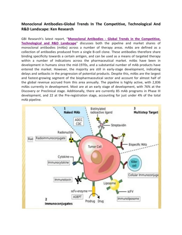
MAb therapy targets Inflammation Autoimmune disease Graft rejection Cancer Viral infection
Therapeutic strategies Plain MAbs MAbs fused to other protein binders (e.g., soluble receptors) to increase avidity and/or to effect ADCC MAbs fused to cytotoxic agents (toxins, radionuclides) Toxins: ricin (stops protein synthesis) calicheamicin (DNA breaks) Radionuclides: 90Y = yttrium 111I = indium
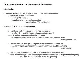
Monoclonal antibody generation • - Cells needed myeloma cells and mouse spleen cells • - antigen administration Kohler and Milstein • - hybridoma formation via cell fusion • selection mutants required (myeloma hprt- usually) • Further development: • - antibody generation cDNA cloning from hybridoma • - engineered MAbs expression vectors • refinement 2nd generation antibodies, in vitro • Solve problems of using mouse antibodies in humans
Problems of mouse MAbs • Fc portion limited in its ability to interact with Fc receptors of human cells. • Lower serum half-life • Development of human anti-mouse antibodies (HAMA) • Retreatment results in allergy or anaphylactic shock • Retreatment is less effective Breedveld, Lancet 2000 355:9205 • Solutions via recombinant DNA genetic engineering : • Chimeric mouse-human antibodies: mouse V regions fused to Hu C-region • Humanized mouse antibodies, Parts of V-region from human interspersed with mouse CDR V-regions • Human antibodies (fully), via transgenic mice carrying human immunoglobulin genes as source of spleen cells (Medarex, Abgenix, Kirin) CDR = complementarity-determining region
MAb Fusion Proteins With other protein-binding proteins: natural receptors in soluble form Analogous to MAbs and make use of the Fc portion of the antibody molecule: Example: Enbrel (etanercept): Anti-rheumatoid arthritis drug Soluble TNF receptor fused to the Fc IgG1 domain (TNF= tumor necrosis factor) Ties up TNF, blocking its inflammatory function Fc domain dimerizes the receptor, which increases its affinity for TNF. Fc domain increases the half-life of the protein in the bloodstream Amgen + Wyeth Example, still experimental: Anti-HIV drug PRO 542 Uses soluble form of the CD4, the molecule to which HIV attaches on T-cells Aim: block the viral surface protein that binds CD4 Soluble CD4 (HIV receptor) fused to IgG2. Tetrameric (all 4 V-regions replaced) – therefore mutlivalent Reduced Fc function (chose IgG2 for this reason) Better half-life than soluble CD4 itself (Recently replaced by a MAb (PRO 140) targeting the CCR5 cell surface protein, required for viral entry) Progenics
Ag binding site 15 AA linker Single chain antibodies (scFv)
Phage display to isolate functional V-regions Can be used to screen billions of V-region variants for binding to a particular antigen of choice. Key requirement of this powerful strategy, and many of a similar kind:A physical link of 1) a nucleic acid sequence (here, DNA) to 2) the phenotype (e.g., binding to something) of a protein coded by that nucleic acid Commonly used phage = m13, filamentous, infects E. coli Phage coat protein the protein the DNA(inside) “Panning” E.g., for a SC Ab, coat the dish with Ag Protein displayed in the phage coatCan screen 108phage
Phage display selection of scFv’s (single-chain variable regions) Source of sequence: PCR from genome or RT-PCR from mRNA, add randomization (doped synthesis). Repeat, to reduce background Wash or Elute, re-infect PCR rescue scFv DNA Clone (plaque)