Download
1 / 34
340 likes | 491 Views
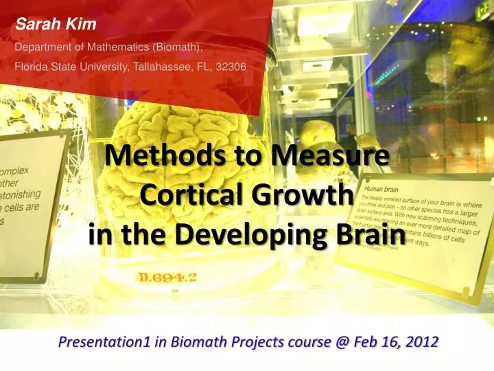
Sarah Kim Department of Mathematics (Biomath), Florida State University, Tallahassee, FL, 32306. Methods to Measure Cortical Growth in the Developing Brain. Presentation1 in Biomath Projects course @ Feb 16, 2012 . We are interested in the folding pattern in the growing brain.

E N D
Sarah Kim Department of Mathematics (Biomath), Florida State University, Tallahassee, FL, 32306 Methods to Measure Cortical Growth in the Developing Brain Presentation1 in Biomath Projects course @ Feb 16, 2012
We are interested in the folding pattern in the growing brain. Human Brain at the Science Museum - London
Gyrus (ridge) Sulcus (valley) Human Brain at the Science Museum London
Folding and Surface Area The cortical folding increases the surface area of the cortex relative to the brain volume, and permits a rapid increase of the cortical surface area during significant growth phases of the cortical volume. (Reference: Richman et al., 1975)
Sarah Kim Department of Mathematics (Biomath), Florida State University, Tallahassee, FL, 32306 Biological Background
Cerebrum & Cerebellum CEREBRUM CEREBELLUM A human brain, with the cerebellum colored in purple.
6 Major Lobes Parietal Lobe Frontal Lobe Occipital Lobe CEREBRUM Insular Lobe Temporal Lobe CEREBELLUM Limbic Lobe
6 Major Lobes Insular Lobe Limbic Lobe Note: The colors of other lobes are not matched with previous figure.
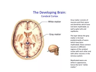
In anatomy, the cortex means the outmost layer of an organ. * Adjective: Cortical Cortex White matter Grey matter The cerebral cortex is the outer layer depicted in dark violet. The cerebellum cortex
Major Sulci Central sulcus Lateral fissure Parietooccipital sulcus Preoccipital notch Cingulate sulcus Reference: www.AnatomyGuy.com Neuroanatomy: Lobes and Major Fissures of the Human Brain
Ventricular System of the Human Brain Right Lateral Ventricle (LV) Left Lateral Ventricle (LV) Third Ventricle Ref: http://biology.about.com/library/organs/brain/blventricles.htm Fourth Ventricle
Ventricular System of the Human Brain Subventricular Zone (SVZ) Proliferative cells Ventricular Zone (VZ) later before Ref: A. Kriegstein et al. (2006) Nature Reviews Neuroscience, 7(11), 883-890
Abnormal Cortical Folding - Lissencephaly • Lissencephaly • → Smooth surfaced brain • with total absence of gyri. • Tulane University School of Medicine Image of LissencephalicBrain UC San Diego School of Medicine
Abnormal Cortical Folding - Polymicrogyria Generalized Polymicrogyria Northeast Ohio Medical University Brain of a Dog died from Polymicrogyria green arrow: normal part red arrow: polymicrogyria part University of Missouri – Columbia College of Veterinary Medicine
Even Normal Cortical Folding Even normal folding is incompletely understood.
Sarah Kim Department of Mathematics (Biomath), Florida State University, Tallahassee, FL, 32306 Research History
Why do we need mathematical models? • The brain is one of nature’s great mysteries. • The folding patterns are unique, both across species and across individuals within a species. • There are neurological disorders that result in abnormal cortical folding. You are special!
Why do we need mathematical models? • The mechanism behind cortical folding is unknown. • There is debate about the mechanism of cortical folding. (Existing biological models of folding conflict) • Difficult to perform neuroscience experiments on cortical folding. Chemical Based Models Mechanical Based Models
Why do we need mathematical models? Because of debate and lack of experiments, the mathematical models are needed to analytically describe cortical folding.
Two Competing Theories Mechanical Based Models Chemical Based Models KriegsteinA et al. (2006) Nat Rev Neurosci 7: 883–890. Patterns of neural stem and progenitor cell division may underlie evolutionary cortical expansion. David Van Essen (1997) Nature 385:313-318 A tension-based theory of morphogenesis and compact wiring in the central nervous system. Deborah A Striegel, Monica K Hurdal (2009) PLoS Computational Biology Chemically Based Mathematical Model for Development of Cerebral Cortical Folding Patterns Gregory Toole, Monica K Hurdal Modeling Cortical Folding Patterns of the Brain Using a Growing Domain
Mechanical Based Models Biomechanismof cortical folding are proposed. 1. Tension Forces applied by white matter fibers Proposed by Van Essen (1997) Modeled based on DTI data by Basser et al. (1994) → the white matter tension and direction 2. Cortical Growth (radial and tangential directions) Modeled based on MRI data by Geng et al. (2009) → the anisotropic growth of the cortex, regulated by inhomogeneous white matter rigidity
Mechanical Based Models Elastic force Plasticity
Mechanical Based Models
Mechanical Based Models
Mechanical Based Models
Kriegstein A et al. (2006) Nat Rev Neurosci 7: 883–890. Patterns of neural stem and progenitor cell division may underlie evolutionary cortical expansion. Chemical Based Models Intermediate Progenitor Model (Kriegstein et al. 2006) implicates Intermediate Progenitor Cells (IPCs) in SVZ in cortical development R N N N N N N N N N Cortical plate - Radial glial cell N - Neuron N N N N N N - Intermediate progenitor cell Intermediate zone SVZ I I VZ R R R Lateral Ventricle (LV) N I
Kriegstein A et al. (2006) Nat Rev Neurosci 7: 883–890. Patterns of neural stem and progenitor cell division may underlie evolutionary cortical expansion. Chemical Based Models later before
Chemical Based Models Turing System Approximation of lateral ventricle with prolatespheroid.
Chemical Based Models
Sarah Kim Department of Mathematics (Biomath), Florida State University, Tallahassee, FL, 32306 Future Work / My Questions
Mechanical + Chemical? How? Only Mechanical? Only Chemical?
Sarah Kim Department of Mathematics (Biomath), Florida State University, Tallahassee, FL, 32306 Question?Comments?
Sarah Kim Department of Mathematics (Biomath), Florida State University, Tallahassee, FL, 32306 Thank you!