Download

1 / 36
360 likes | 474 Views
Mediators of vascular remodelling co-opted for sequential steps in lung metastasis. Gaorav P. Gupta1*, Don X. Nguyen1*, Anne C. Chiang1,2, Paula D. Bos1, Juliet Y. Kim1, Cristina Nadal1, Roger R. Gomis1, Katia Manova-Todorova3 & Joan Massague´1,4. Goal and strategy. Main goal:

E N D
Mediators of vascular remodelling co-opted for sequential steps in lung metastasis Gaorav P. Gupta1*, Don X. Nguyen1*, Anne C. Chiang1,2, Paula D. Bos1, Juliet Y. Kim1, Cristina Nadal1, Roger R. Gomis1, Katia Manova-Todorova3 & Joan Massague´1,4
Goal and strategy Main goal: To test if a group of genes contribute collectively to angiogenesis in mammary tumours, the entry of tumour cells into the circulation and their exit from the blood stream into the pulmonary parenchyma. Strategy: Based on previous studies1, four genes were chosen for further investigations: • EREG (pan-HER ligand epiregulin) • COX2 (cyclooxygenase 2) • MMP1 (Matrix metalloproteinase 1) • MMP2 (Matrix metalloproteinase 2) 1Minn, A. J. et al. Genes that mediate breast cancer metastasis to lung. Nature 436, 518–524 (2005). Minn, A. J. et al. Lung metastasis genes couple breast tumor size and metastatic spread. Proc. Natl Acad. Sci. USA 104, 6740–6745 (2007).
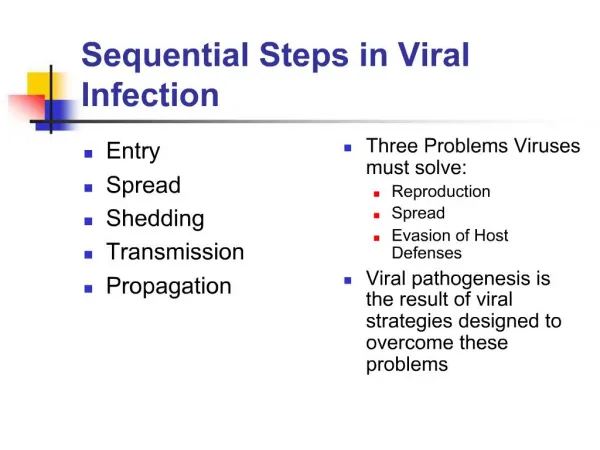
Primary tumour Proliferation Apoptosis Healthy cells Blood vessel Secondary tumour site Metastasis formation Invasive tumour cell Intravasation Tumour cell in the blood vessel Tumour cell in the blood vessel wall Extravasation Metastases genesis
Genes products: EREG • FUNCTION: May be a mediator of localized cell proliferation. As a mitogen it may stimulate cell proliferation and/or angiogenesis. • SUBCELLULAR LOCATION: Precursor form: Cell membrane; Single-pass type I membrane protein. Mature form: Secreted, extracellular space. • TISSUE SPECIFICITY: In normal adults, expressed predominantly in the placenta and peripheral blood leukocytes. High levels were detected in carcinomas of the bladder, lung, kidney and colon. • SIMILARITY: Contains 1 EGF-like domain.
Genes products: COX2 • FUNCTION: May have a role as a major mediator of inflammation and/or a role for prostanoid signaling in activity-dependent plasticity. • CATALYTIC ACTIVITY: Arachidonate + AH2 + 2 O2 = prostaglandin H2 + A + H2O. • PATHWAY: Fatty acids biosynthesis; prostaglandin biosynthesis. • SUBCELLULAR LOCATION: Microsome membrane; Peripheral membrane protein. • INDUCTION: By cytokines and mitogens. • MISCELLANEOUS: This enzyme is the target of nonsteroidal anti-inflammatory drugs such as aspirin. Prostaglandins: lipid compounds that have paracrine or autocrine effects. Potent but with short half-life. • Cause constriction or dilatation in vascular smooth muscle cells, as well as permeability changes • Cause aggregation or disaggregation of platelets • Sensitize spinal neurons to pain • Constrict smooth muscle • Regulate inflammatory mediation • Regulate calcium movement • Regulate hormone regulation • Control cell growth
Genes products: MMP1 • FUNCTION: Cleaves collagens of types I, II, and III at one site in the helical domain. Also cleaves collagens of types VII and X. • CATALYTIC ACTIVITY: Cleavage of the triple helix of collagen at about three-quarters of the length of the molecule from the N-terminus, at 775-Gly-|-Ile-776 in the alpha-1(I) chain. Cleaves synthetic substrates and alpha-macroglobulins at bonds where P1' is a hydrophobic residue. • SUBCELLULAR LOCATION: Secreted. • DOMAIN: There are two distinct domains in this protein; the catalytic N-terminal, and the C-terminal which is involved in substrate specificity and in binding TIMP (tissue inhibitor of metalloproteinases).
Genes products: MMP2 • FUNCTION: In addition to gelatin and collagens, it cleaves KiSS1 at a Gly-|-Leu bond. • CATALYTIC ACTIVITY: Cleavage of gelatin type I and collagen types IV, V, VII, X. Cleaves the collagen-like sequence Pro-Gln-Gly-|-Ile-Ala-Gly-Gln. • TISSUE SPECIFICITY: Produced by normal skin fibroblasts. • PTM: The propeptide is processed by MMP14 (MT-MMP1) and MMP16 (MT-MMP3).
MMP’s • Matrix metalloproteinases (MMPs) are zinc-dependent endopeptidases • There are currently 28 known human MMPs family members • Collectively they are capable of degrading all kinds of extracellular matrix proteins, but also can process a number of bioactive molecules. They are known to be involved in the cleavage of cell surface receptors, the release of apoptotic ligands (such as the FAS ligand), and chemokine in/activation. MMPs are also thought to play a major role on cell behaviors such as cell proliferation, migration (adhesion/dispersion), differentiation, angiogenesis, apoptosis and host defense.
Methods: Generation of cell clones • LM2-4175 • Subpopulation of MDA-MB-231 lung metastatic • Knockdown: with short hairpin RNA interference (shRNA) • Overexpression: by plasmid infection • Immunocompromided mices
Strategy • Working with cells • Primary tumour • Reducing expression of EREG, COX2, MMP1 & 2, inoculation in mice fat pad to check primary tumour growth • Overexpression of EREG, COX2, MMP1 & 2 in knockdown cells, inoculation in mice fat pad to check primary tumour growth • Vessels study • Metastases • Knockdown cells inoculation in vein to assess the metastatic lung progression • Overexpressed cells inoculation in vein to assess the metastatic lung progression • Working with drugs • Inoculation of LM2 cells in mouse fat pad • Analysis of primary tumour • Analysis of intravasation/extravasation • Analysis on metastases • Tests with cells from fresh tissues • Results with drugs provision
Tumour cells growth - knockdown cells • Injections of knockdown cells into mice mammary fat pad • Individually gene knockdown has limited but statistically significant effects on tumour growth. • Silencing all four genes simultaneously arise a nearly complete abrogation of growth. asterisk, P<0.05; double asterisk, P<0.01; triple asterisk, P<0.001; calculated using a two-tailed Student’s t-test for tumour volumes at the last time point, compared to control.
Tumour cells growth - rescue cells • Overexpressing MMP1, MMP2 and COX2 resulted in significant recovery of lung metastatic activity
Why? Proliferation? • Quantified phosphorylated histone H3 levels indicates that the proliferation rate was not significantly alterated.
Why? Apoptosis? • An increased rate of apoptosis was evident in tumours with combinatorial knockdown cells. single asterisk, P < 0.01; double asterisk, P < 0.001
Why? Hypothesis: • Increased rates of tumour cells death might be secondary to defects in angiogenesis. Let’s check…
Why? Checking vessels structures… • Vessels structure Control 4-shRNA Stained with CD31 antibody
Control 4-shRNA Why? Checking vessels permeability… • Vessels permeability Injection of rhodamine-conjugated dextran
Why? Checking pericyte recruitment… • Further analysis did not reveal any major differences in pericyte recruitment.
Why? Checking VEGF… • These defects in primary tumour vessels morphology occurred in the absence of differences in VEGF levels between control and knockdown tumour cells.
Why? Conclusions • No major differences in cells proliferation rate • Increased rate of apoptosis • No considerably alteration on discrete vessel units • No major differences on pericyte recruitment • No differences on VEGF levels • But: reducing of length, number of lumens and extent of branching • This suggests that the 4 genes in tumour tissues promote the formation of dilated, tortuous and leaky blood vessels that typify the neovasculature of aggressive primary tumours.
Control 4-shRNA Pulmonary metastases study • Injections of knockdown cells intravenously into mice • Monitoring by bioluminescence • Histological examination
Extravasation from lung capillaries: Control Red: rhodamine-conjugated lectin injected 48 h. after cells inoculation. Green:tumor cells with cell tracker green
Migratory capacity: in vitro assay • Confirm the inhibition of migratory capacity of LM2 4-shRNA cells through an endotelial monolayer • This do not entail defect in cell motility NoEC – No endothelial monolayer HUVEC – Human umbilical vein endothelial cell HPMEC – Human pulmonary microvascular endothelial cell
Conclusions • The results provide evidence that the expression of ERG, COX2, MMP1 & 2 by cancer cells can collectively promote metastatic extravasation in the lungs.
Inhibition by drugs • Cetuximab: anti-EGFR antibody • Celecoxib: COX2 inhibitor • GM6001: broad-spectrum MMP inhibitor Reported effects on primary tumour growth • Inhibition of tumour growth • Vascular defects -> hypoxia & apoptosis • Impaired tumour cells intravasation from the primary site • Impaired tumour cells extravasation from blood vessels
Inhibition of primary tumour growth • Single agent: minimally inhibition or tumour growth • 3-combined: reduced rate of tumour growth
Effects on intravasation • Presence of circulating tumour cells was assessed by measuring the relative expression of human-specific GAPDH in blood from treated mice.
Control Drug Effects on extravasation
Effects on colonization inhibition • Mice pre-treated with drugs two days before inoculation • Treatment maintained after inoculation
Effects on colonization inhibition (cont.) • After day 28, drugs treatment was terminated. • Rescue of metastases (Off-drug pict.) Immunofluorescent detection of tumour-specific vimentin (green) and CD31 (red)
Effects on metastases size Asterisk, P<0.05; double asterisk, P<0.01; NS, not significant, based on a two-tailed Wilcoxon rank-sum test.
Notes • An exception to this trend was the antagonistic interaction between celecoxib and GM6001, perhaps reflecting the fact that the latter is a broad-spectrum MMP inhibitor, likely to affect both pro- and anti-metastatic MMPs
Moreover • Similar experiments was performed with fresh malignant cells obtained from the pleural fluid of two patients with advanced breast cancer and a diagnosis of lung metastasis. • A subpopulation (CN34.2A) that expresses high levels of EREG, COX2 and MMP1 was inoculated in mice, arising an elevated lung colonization, which could be inhibited by administration of cetuximab and celecoxib. • A sample from a different patient (CN41) was used without prior selection of metastatic cells. They had basal lung colonizing activity that could also be inhibited by the celecoxib/cetuximab combination.
Paper discussion • What is the key experiment? (the one confirming the statement in the title) • What is the strongest point? • What is the weakest point? and What to do to strenghten it? • What is the take home message? (summarize it in a sentence)