Download
1 / 27
290 likes | 686 Views
Chapter 4 DNA, RNA and the Flow of Genetic Information. Backbone. Phosphodiester bond (-) charges protect “P” from being attacked by nucleophiles . Absence of 2’ –OH in DNA increase its resistance to hydrolysis Histones provide (+) charges for neutralization. Nucleic Acids.

E N D
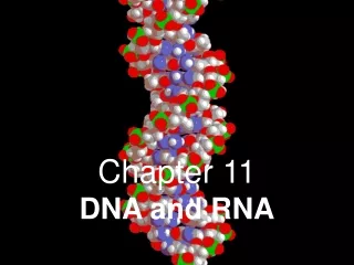
Chapter 4 DNA, RNA and the Flow of Genetic Information
Backbone • Phosphodiester bond • (-) charges protect “P” from being attacked by nucleophiles. • Absence of 2’ –OH in DNA increase its resistance to hydrolysis • Histones provide (+) charges for neutralization. Nucleic Acids • DNA and RNA are unbranched linear polymers built up from similar units. • Each monomer unit within the polymer consists of three components : a sugar, a phosphate, and a base
A, G, C, T for DNA A, G, C, U for RNA Bases : Purines & Pyrimidines linkage with sugar through N9 linkage with sugar through N1
Sugar-Base Linkages between C1-OH of Sugars and N9 of Purines or C1-OH of Sugars and N1 of Pyrimidines Sugar-Phosphate Linkages between C3-OH of Sugar and Phosphate or C5-OH of Sugar and Phosphate Nucleoside Nucleoside Phosphate; Nucleotide
Polarity of DNA Chain • 5’ end OH is usually occupied by a phosphate group. • 3’ end OH is usually open as unmodified. • By convention, DNA base sequence is 5’ 3’ direction. • pACG ≠ pGCA
DNA Double Helix : Watson & Crick • Right-handed coiled helix with two polynucleotide chains • Base : Inside • Sugar & Phosphate : Outside • Bases are perpendicular to the helical axis. • Adjacent bases separated by 3.4Å • The same helical structure repeats every 34Å. • 10 bases per turn of helix • Rotation of 36 degree per base • Diameter of the helix : 20Å • Hydrophobic interactions between bases inside; hydrophilic polar surface
Watson-Crick Base Pairing in DNA by Hydrogen Bondings 1-5 kcalmol-1 Chargaff’s rule
Stacking of Base pairs for stability • Hydrophobic effect just like protein • van der Waals forces - (0.5-1 kcalmol-1) The Double heix facilitates the accurate transmission of Hereditary information Semiconservative replication test by Dr. Meselson and Dr. stahl
Genomic DNA Labeling with 15NH4Cl Density Gradient Centrifugation with CsCl Complementary & Semi-Conservative DNA Replication
Hypochromism by Base Pairing Denaturation of DNA by heat, acid or alkali DNA sequence similarity between different organisms (i.e. relatedness) can be determined by the degree of hybridization. Tm : the temperature at which half the helical structure is lost Melting & Annealing of DNA
Linearity of DNA Circular DNA vs. Linear DNA Prokaryotic vs. Eukaryotic Mitochondrial vs. Genomic DNA Supercoiling Helix vs. Superhelix Topoisomerase Structural Stability of Cellular DNA Regulation of Gene Expression DNA in vivo Packing Fold : 1000 times Relaxed Circular DNA Supercoiled Circular DNA DNA Topology
Stem-Loop (Hairpin) Unusual Base Pairing between Three Bases (Long Range Interaction) Complex Structures Formed by Single Strand Nucleic Acids
Replication by DNA Polymerase • Take instructions from templates (pre-existing DNA strands) • DNA synthesis through complementary base pairing • Step-by-step addition of deoxyribonucleotide to a DNA chain • (DNA)n + 5’-dNTP (DNA)n+1 + PPi • Template • Primer strand with a free 3’-OH group • Nucleotides : dATP, dGTP, dCTP, TTP • Divalent metal ion : Mg2+
DNA Polymerase Reaction • Complementary Base Pairing between template and incoming dNTP • (DNA Polymerase : Template-Directed Enzyme) • 2. Nucleophilic attack by 3’-OH of the primer strand on the inner most phosphorous atom of the incoming dNTP • Subsequent pyrophosphate (PPi) hydrolysis by pyrophosphatase provides further driving force for the reaction. • DNA elongation direction : 5’ 3’ • Exonuclease activity by DNA polymerase removes mismatched bases during synthesis (3’5’) and after synthesis (5’3’) : proof-reading • (Error rate of DNA polymerase = 10-8 per base pair
Some Viral Genomes Are Made of RNA RNA Virus single-stranded RNA (viral genome) Protein RNA-directed RNA polymerase for RNA replication (e.g. Tobacco Mosaic Virus) Retro-Virus single-stranded RNA (viral genome) RNA-directed DNA synthesis by reverse transcriptase single-stranded DNA double-stranded DNA integrate into the host genome replication together with the host genome later, when it is necessary, express viral RNA and proteins packaging virus particles and exit from the host (e.g. HIV-1)
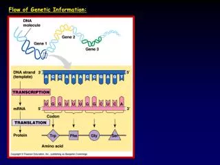
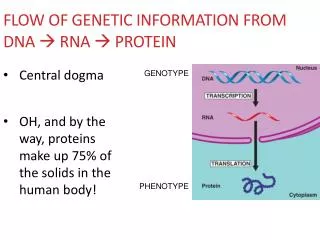
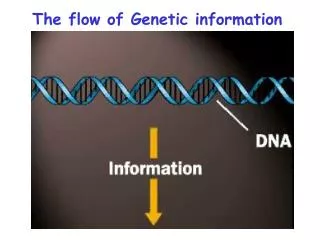
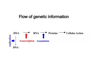
Central Dogma : DNA RNA Protein DNA ▪ storage of genetic information ▪ serve only as information source during gene expression processes ▪ minimize the chances of mutation RNA ▪ photocopy of genetic information from DNA ▪ dictate the repertoire of the proteins to be expressed (mRNA) ▪ exist transiently as multiple copies (mRNA) ▪ assist protein translation (rRNA, tRNA) ▪ assist mRNA splicing and nuclear export (snRNA, hnRNA)
RNA Polymerase Reaction • Complementary Base Pairing between DNA template and incoming NTP • (RNA Polymerase : DNA-Directed RNA Synthesizing Enzyme) • Step-by-step addition of ribonucleotide to the RNA primer strand • (RNA)n + 5’-NTP (RNA)n+1 + PPi ; Primer is NOT required ; • Nucleotides : ATP, GTP, CTP, UTP ; Divalent metal ion : Mg2+, Mn2+ • Nucleophilic attack by 3’-OH on the phosphorous atom of the incoming NTP • Subsequent pyrophosphate (PPi) hydrolysis • RNA elongation direction : 5’ 3’ ; No proof-reading function
RNA Polymerases Take Instructions from DNA Templates • Base Composition (Viral DNA vs. Viral RNA) • Hybridization Experiments between DNA template and transcribed RNA • Sequence Comparison between RNA and DNA templates • (template strand vs. coding strand; anti-sense strand vs. sense strand)
Promoter (Transcriptional Initiation) • Binding Sites for RNA Polymerase for Transcriptional Initiation • cf. TBP (TATA Binding Protein); TAFs (TBP Associated Factors); Basal Machinery • Binding Sites for Various Transcription Factors for More Complex Regulation of Transcription (Enhancer)
Terminator (Transcriptional Termination) • Hairpin Forming Sequences • Poly-U Stretches • Transcription Termination Protein : rho Post-Transcriptional Modification of mRNA 5’ Capping & 3’ Poly-Adenylation
Transfer RNAs Bring Amino Acids to the mRNA Template during Translation (Protein Synthesis by Ribosomes) tRNA charged with amino acid : aminoacyl-tRNA : aa-tRNA
Triplet Codons Specify Each Amino Acid • Three nucleotides encode an amino acid. • The code is non-overlapping. • The code is sequentially translated. • The genetic code is degenerated. • 43 = 64 = 61 coding codons + 3 stop codons • Codon degeneracy decreases the chances for translational termination (64 = 20 + 44 ?). • Codon degeneracy also reduces protein sequence changes by genetic mutations. • Codon degeneracy is most often found in Wobble position (3rd base in a triplet codon) • Recognition of stop codons by release factor
Conjugated with Initiator tRNA mRNA Contains Start and Stop Signals for Translation • The first AUG (or GUG) is recognized by fMet-tRNA during translational initiation. • Internal AUGs are recognized by Met-tRNA, and GUGs are by Val-tRNA. • IF (initiation factor) vs. EF (elongation factor) for interactions with aa-tRNA • Location of initiator AUG determines the reading frame for following triplet codons.
The Genetic Code Is Nearly Universal • The codon usage is almost invariant throughout the evolution. • Translation of mRNAs from foreign species is usually successful. • BUT, codon preference differs quite a bit between organisms. • Mitochondrial gene expression utilizes slightly different codons.
Exon: segments of nascent mRNA retained in the mature mRNA (usu. coding one domain) Intron: segments of nascent mRNA absent in the mature mRNA Detection of single-stranded DNA upon DNA-mRNA hybridization (electron microscopy) Splicing Typical Intron Structure Mosaic Nature of Eukaryotic Genes : Introns & Exons
Generation of Novel Genes by Exon Shuffling during Evolution Alternative Splicing Allows Generation of Protein Variants from One Gene