Download
1 / 40
400 likes | 588 Views
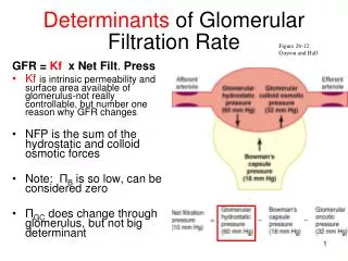
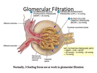
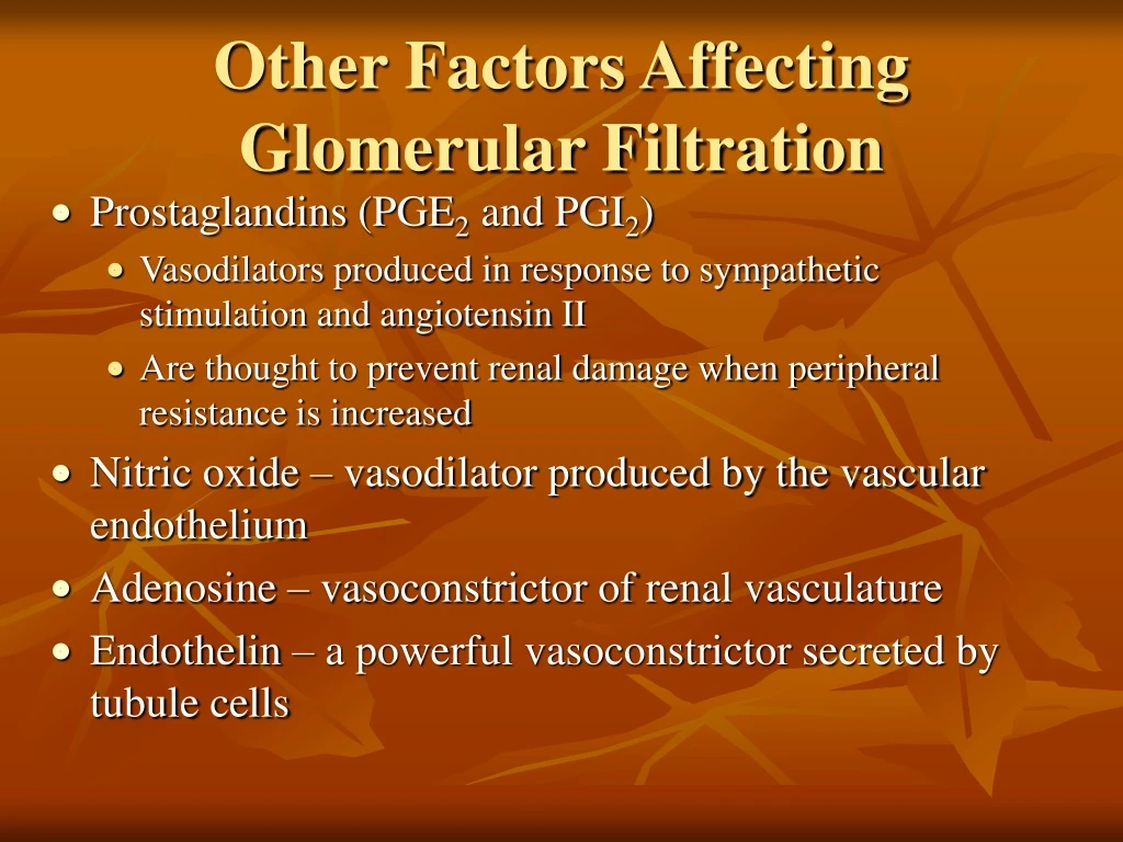
Other Factors Affecting Glomerular Filtration. Prostaglandins (PGE 2 and PGI 2 ) Vasodilators produced in response to sympathetic stimulation and angiotensin II Are thought to prevent renal damage when peripheral resistance is increased

E N D
Other Factors Affecting Glomerular Filtration • Prostaglandins (PGE2 and PGI2) • Vasodilators produced in response to sympathetic stimulation and angiotensin II • Are thought to prevent renal damage when peripheral resistance is increased • Nitric oxide – vasodilator produced by the vascular endothelium • Adenosine – vasoconstrictor of renal vasculature • Endothelin – a powerful vasoconstrictor secreted by tubule cells
Tubular Reabsorption • A transepithelial process whereby most tubule contents are returned to the blood • Transported substances move through three membranes • Luminal and basolateral membranes of tubule cells • Endothelium of peritubular capillaries • Only Ca2+, Mg2+, K+, and some Na+ are reabsorbed via paracellular pathways
Tubular Reabsorption • All organic nutrients are reabsorbed • Water and ion reabsorption is hormonally controlled • Reabsorption may be an active (requiring ATP) or passive process
Sodium Reabsorption: Primary Active Transport • Sodium reabsorption is almost always by active transport • Na+ enters the tubule cells at the luminal membrane • Is actively transported out of the tubules by a Na+-K+ ATPase pump
Sodium Reabsorption: Primary Active Transport • From there it moves to peritubular capillaries due to: • Low hydrostatic pressure • High osmotic pressure of the blood • Na+ reabsorption provides the energy and the means for reabsorbing most other solutes
Reabsorption by PCT Cells • Active pumping of Na+ drives reabsorption of: • Water by osmosis, aided by water-filled pores called aquaporins • Cations and fat-soluble substances by diffusion • Organic nutrients and selected cations by secondary active transport
Nonreabsorbed Substances • A transport maximum (Tm): • Reflects the number of carriers in the renal tubules available • Exists for nearly every substance that is actively reabsorbed • When the carriers are saturated, excess of that substance is excreted
Nonreabsorbed Substances • Substances are not reabsorbed if they: • Lack carriers • Are not lipid soluble • Are too large to pass through membrane pores • Urea, creatinine, and uric acid are the most important nonreabsorbed substances
Absorptive Capabilities of Renal Tubules and Collecting Ducts • Substances reabsorbed in PCT include: • Sodium, all nutrients, cations, anions, and water • Urea and lipid-soluble solutes • Small proteins • Loop of Henle reabsorbs: • H2O, Na+, Cl, K+ in the descending limb • Ca2+, Mg2+, and Na+ in the ascending limb
Absorptive Capabilities of Renal Tubules and Collecting Ducts • DCT absorbs: • Ca2+, Na+, H+, K+, and water • HCO3 and Cl • Collecting duct absorbs: • Water and urea
Na+ Entry into Tubule Cells • Passive entry: Na+-K+ ATPase pump • In the PCT: facilitated diffusion using symport and antiport carriers • In the ascending loop of Henle: facilitated diffusion via Na+-K+-2Cl symport system • In the DCT: Na+-Cl– symporter • In collecting tubules: diffusion through membrane pores
Atrial Natriuretic Peptide Activity • ANP reduces blood Na+ which: • Decreases blood volume • Lowers blood pressure • ANP lowers blood Na+ by: • Acting directly on medullary ducts to inhibit Na+ reabsorption • Counteracting the effects of angiotensin II • Indirectly stimulating an increase in GFR reducing water reabsorption
Tubular Secretion • Essentially reabsorption in reverse, where substances move from peritubular capillaries or tubule cells into filtrate • Tubular secretion is important for: • Disposing of substances not already in the filtrate • Eliminating undesirable substances such as urea and uric acid • Ridding the body of excess potassium ions • Controlling blood pH
Regulation of Urine Concentration and Volume • Osmolality • The number of solute particles dissolved in 1L of water • Reflects the solution’s ability to cause osmosis • Body fluids are measured in milliosmols (mOsm) • The kidneys keep the solute load of body fluids constant at about 300 mOsm • This is accomplished by the countercurrent mechanism
Countercurrent Mechanism • Interaction between the flow of filtrate through the loop of Henle (countercurrent multiplier) and the flow of blood through the vasa recta blood vessels (countercurrent exchanger) • The solute concentration in the loop of Henle ranges from 300 mOsm to 1200 mOsm • Dissipation of the medullary osmotic gradient is prevented because the blood in the vasa recta equilibrates with the interstitial fluid
Loop of Henle: Countercurrent Multiplier • The descending loop of Henle: • Is relatively impermeable to solutes • Is permeable to water • The ascending loop of Henle: • Is permeable to solutes • Is impermeable to water • Collecting ducts in the deep medullary regions are permeable to urea
Loop of Henle: Countercurrent Exchanger • The vasa recta is a countercurrent exchanger that: • Maintains the osmotic gradient • Delivers blood to the cells in the area
Formation of Dilute Urine • Filtrate is diluted in the ascending loop of Henle • Dilute urine is created by allowing this filtrate to continue into the renal pelvis • This will happen as long as antidiuretic hormone (ADH) is not being secreted
Formation of Dilute Urine • Collecting ducts remain impermeable to water; no further water reabsorption occurs • Sodium and selected ions can be removed by active and passive mechanisms • Urine osmolality can be as low as 50 mOsm (one-sixth that of plasma)
Formation of Concentrated Urine • Antidiuretic hormone (ADH) inhibits diuresis • This equalizes the osmolality of the filtrate and the interstitial fluid • In the presence of ADH, 99% of the water in filtrate is reabsorbed
Formation of Concentrated Urine • ADH-dependent water reabsorption is called facultative water reabsorption • ADH is the signal to produce concentrated urine • The kidneys’ ability to respond depends upon the high medullary osmotic gradient
Diuretics • Chemicals that enhance the urinary output include: • Any substance not reabsorbed • Substances that exceed the ability of the renal tubules to reabsorb it • Substances that inhibit Na+ reabsorption
Diuretics • Osmotic diuretics include: • High glucose levels – carries water out with the glucose • Alcohol – inhibits the release of ADH • Caffeine and most diuretic drugs – inhibit sodium ion reabsorption • Lasix and Diuril – inhibit Na+-associated symporters
Renal Clearance • The volume of plasma that is cleared of a particular substance in a given time • Renal clearance tests are used to: • Determine the GFR • Detect glomerular damage • Follow the progress of diagnosed renal disease
Renal Clearance RC = UV/P RC = renal clearance rate U = concentration (mg/ml) of the substance in urine V = flow rate of urine formation (ml/min) P = concentration of the same substance in plasma
Physical Characteristics of Urine • Color and transparency • Clear, pale to deep yellow (due to urochrome) • Concentrated urine has a deeper yellow color • Drugs, vitamin supplements, and diet can change the color of urine • Cloudy urine may indicate infection of the urinary tract
Physical Characteristics of Urine • Odor • Fresh urine is slightly aromatic • Standing urine develops an ammonia odor • Some drugs and vegetables (asparagus) alter the usual odor
Physical Characteristics of Urine • pH • Slightly acidic (pH 6) with a range of 4.5 to 8.0 • Diet can alter pH • Specific gravity • Ranges from 1.001 to 1.035 • Is dependent on solute concentration
Chemical Composition of Urine • Urine is 95% water and 5% solutes • Nitrogenous wastes: urea, uric acid, and creatinine • Other normal solutes include: • Sodium, potassium, phosphate, and sulfate ions • Calcium, magnesium, and bicarbonate ions • Abnormally high concentrations of any urinary constituents may indicate pathology
Ureters • Slender tubes that convey urine from the kidneys to the bladder • Ureters enter the base of the bladder through the posterior wall • This closes their distal ends as bladder pressure increases and prevents backflow of urine into the ureters
Ureters • Ureters have a trilayered wall • Transitional epithelial mucosa • Smooth muscle muscularis • Fibrous connective tissue adventitia • Ureters actively propel urine to the bladder via response to smooth muscle stretch
Urinary Bladder • Smooth, collapsible, muscular sac that stores urine • It lies retroperitoneally on the pelvic floor posterior to the pubic symphysis • Males – prostate gland surrounds the neck inferiorly • Females – anterior to the vagina and uterus • Trigone – triangular area outlined by the openings for the ureters and the urethra • Clinically important because infections tend to persist in this region
Urinary Bladder • The bladder wall has three layers • Transitional epithelial mucosa • A thick muscular layer • A fibrous adventitia • The bladder is distensible and collapses when empty • As urine accumulates, the bladder expands without significant rise in internal pressure
Urethra • Muscular tube that: • Drains urine from the bladder • Conveys it out of the body
Urethra • Sphincters keep the urethra closed when urine is not being passed • Internal urethral sphincter – involuntary sphincter at the bladder-urethra junction • External urethral sphincter – voluntary sphincter surrounding the urethra as it passes through the urogenital diaphragm • Levator ani muscle – voluntary urethral sphincter
Urethra • The female urethra is tightly bound to the anterior vaginal wall • Its external opening lies anterior to the vaginal opening and posterior to the clitoris • The male urethra has three named regions • Prostatic urethra – runs within the prostate gland • Membranous urethra – runs through the urogenital diaphragm • Spongy (penile) urethra – passes through the penis and opens via the external urethral orifice
Micturition (Voiding or Urination) • The act of emptying the bladder • Distension of bladder walls initiates spinal reflexes that: • Stimulate contraction of the external urethral sphincter • Inhibit the detrusor muscle and internal sphincter (temporarily) • Voiding reflexes: • Stimulate the detrusor muscle to contract • Inhibit the internal and external sphincters
Developmental Aspects • Three sets of embryonic kidneys develop, with only the last set persisting • The pronephros never functions but its pronephric duct persists and connects to the cloaca • The mesonephros claims this duct and it becomes the mesonephric duct • The final metanephros develop by the fifth week and develop into adult kidneys
Developmental Aspects • Metanephros develop as ureteric buds that incline mesoderm to form nephrons • Distal ends of ureteric tubes form the renal pelves, calyces, and collecting ducts • Proximal ends called ureteric ducts become the ureters • Metanephric kidneys are excreting urine by the third month • The cloaca eventually develops into the rectum and anal canal
Developmental Aspects • Infants have small bladders and the kidneys cannot concentrate urine, resulting in frequent micturition • Control of the voluntary urethral sphincter develops with the nervous system • E. coli bacteria account for 80% of all urinary tract infections • Sexually transmitted diseases can also inflame the urinary tract • Kidney function declines with age, with many elderly becoming incontinent