Download
1 / 19
190 likes | 362 Views
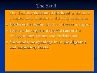
Skull. Anna L. Kiss Department of Anatomy , Histology and Embryology Semmelweis University 2018. Skull : Cranium Mandible Cranial cavity : Roof of the skull : skull cap ( calvaria ) Floor of the skull : base ( basis cranii ). Skull : Neurocranium : frontal parietal

E N D
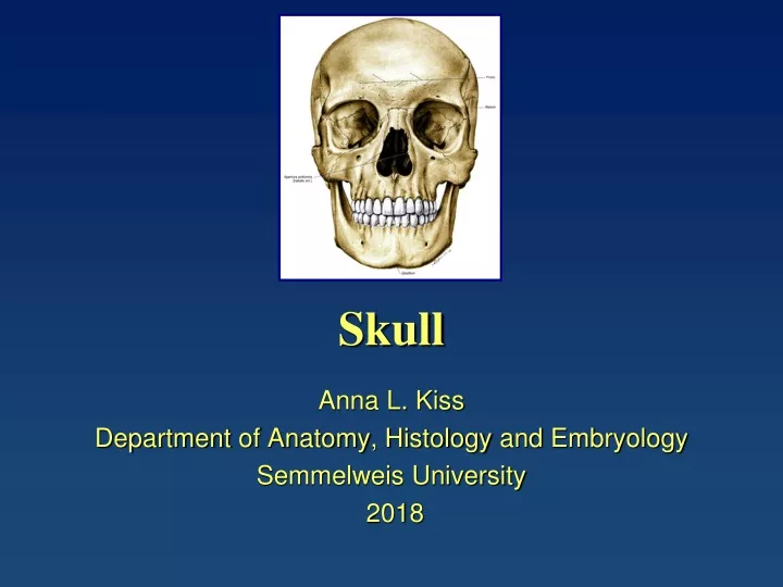
Skull Anna L. Kiss Department of Anatomy, Histology and Embryology Semmelweis University 2018
Skull: • Cranium • Mandible Cranialcavity: Roof of theskull: skullcap (calvaria) Floor of theskull: base (basiscranii)
Skull: Neurocranium:frontal parietal temporal occipital sphenoidal Viscerocranium:maxilla mandible ethmoidal zygomatic nasal lacrimal palatine vomer inf. nasalconcha
Temporalbone squamous part mastoid part
Temporal bone squamous part petrosal part mastoid part
Sphenoidal bone lesser wing greater wing body: sella turcica pterygoid process
Ethmoidal bone Cribriformeplate
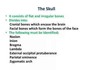
Sutures: (syndesmosis) • coronal: frontal and parietal • sagittal:parietal • lambdoid:parietal and occipital • parieto-mastoid:parietal and temporal • occipito-mastoid:occipital and temporal
Fontanelles: (fonticulus) • anterior • posterior • anterolateral (sphenoidal) • posterolateral (mastoid)
Bregma:middline meeting place of the bones • Pterion:lateral meeting point (parietal, frontal, squama of the temporal, greater wing of sphenoidal) • Asterion:mastoid region meets the occipital and parietal bones
Base: • Anterior cranial fossa:frontal bones (pars orbitalis)+lesser wing of the sphenoidal bone • Middle cranial fossa: from lesser wing of sphenoid bone to pars petrosa of the temporal bone (groove for the sup. petrosal sinus) • Posterior cranial fossa: posterior surface of the pars petrosa (termporal bone)+occipital bone (squama)
Anteriorcranial fossa • Cristagalli (ethmoidalbone) • Cribriformeplate (ethmoidalbone) • Middlecranial fossa • sellaturcica (hypophyseal fossa): sphenoidalbone • sup. orbitalfissure (orbit) • foramenrotundum (fossa pterygopalatina) • foramenovale (externalsurface of theskull) • foramenspinosum(meningealartery) • sulcus n. petrosus major et minor • opticcanal • Posteriorcranial fossa • clivus • foramenmagnum • foramenjugulare • sulcus sinus sigmoideus • sulcus sinus transversus
Skull: inferior view palatine process of the maxilla palatine bone zygomatic arch sphenoidal bone: pterygoid process choana mandibular fossa external opening of the carotid canal foramen magnum jugular foramen and fossa occipital condyle
Hard palate: palatine process of the maxilla+horizontal plate of the palatine bone • Choana: horizontal plate of the palatine bone+pterygoid process of the sphenoidal bone+body of the sphenoidal bone • Atlanto-occipital joint: occipital condyles+sup. art. surfaces of the atlas
References Wikipaedia Sobotta Atlas