Download
1 / 59
600 likes | 857 Views
SYNAPTIC TRANSMISSION I Tim Murphy NRSC 500, 2011 The definition of synaptic transmission is simply the communication between two nerve cells. Communication believed to involve specialized structures termed "synapses". We will focus on: 1) The discovery of synaptic transmission

E N D
SYNAPTIC TRANSMISSION I Tim Murphy NRSC 500, 2011 The definition of synaptic transmission is simply the communication between two nerve cells. Communication believed to involve specialized structures termed "synapses". We will focus on: 1) The discovery of synaptic transmission 2) The form of transmission, i.e. chemical or electrical 3) Criteria for a chemical transmitter 4) Ionic requirement for release 5) Quantal aspects of release: vesicle theory
Discovery of synaptic transmission • Cajal's golgi staining methods suggested the presence of contacts between cells that were used for communication ~1900’s. • Sherrington proposed the term "synapse" meaning to clasp to describe the structure, 1890’s.
Cajal’s drawings Sherrington’s insights 1890s. of golgi stain. Shepherd 1997 TINS
Modern golgi staining, YFP mouse cortical fluorescence, can be bred to other KO’s, transgenics Feng et al. 2000.
From Kristin Harris Lectures. http://synapses.mcg.edu/lab/harris/lectures.htm
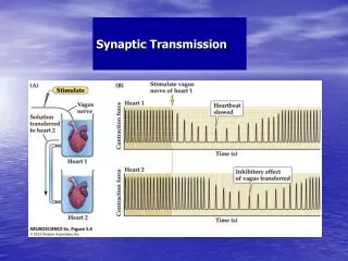
Otto Loewi, chemical transmitter. • 1936 Nobel prize for Medicine • Showed that vagus nerve stimulation liberates a diffusible transmitter. • Perfusate from one stimulated frog heart could be transferred to another and have an effect on beat frequency.
Debate on synaptic transmission - chemical or electrical. • Otto Loewi showed that acetylcholine could mimic the effect of vagal nerve stimulation. • What additional experiment would have been nice to prove that the vagal nerve released acetylcholine. • The results of Loewi's experiments sparked debate about whether chemical and electrical transmission was occurring. • Subsequently shown that both chemical and electrical transmission exist.
From Kristin Harris Lectures. http://synapses.mcg.edu/lab/harris/lectures.htm
Chemical Electrical From Kristin Harris Lectures. http://synapses.mcg.edu/lab/harris/lectures.htm
From Kristin Harris Lectures. http://synapses.mcg.edu/lab/harris/lectures.htm
Electrical Synapses • Gap junction-type communication important for rapidly synchronizing syncytia of cells as is observed in astrocytes, heart, and developing brains. Present in some invertebrates to promote rapid defensive secretions. • Problems with electrical:difficult to modulate gating of channels (exceptions exist cAMP, pH). • Can't change sign, i.e. charge always flows "down hill." • Electrical synaptic transmission requires that the presynaptic cell or terminal be larger than the postsynaptic cell for it to inject considerable charge, no real amplification mechanism.
Electrical synaptic transmission. Fundamental Neuro.
Chemical transmission inhibitors do not block transmission in developing Zebrafish. Saint-Amant and Drapeau Neuron 2001
Gap junction inhibitors block transmission in developing Zebrafish. Saint-Amant and Drapeau Neuron 2001
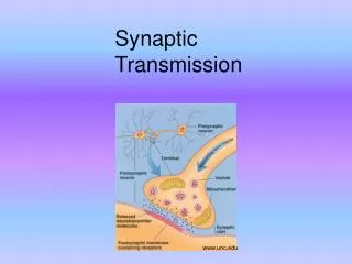
Chemical transmission. • Contrary to electrical transmission multiple steps are required to release transmitter chemicals and for them to act on postsynaptic receptors, resulting in a time delay (can be as short as 0.2 msec, from Ca2+ entry to secretion). • Directional, select localization of release machinery to presynaptic terminals and receptors to postsynaptic specializations. • Can change sign by release of inhibitory transmitter. • Highly modulatable as it has many steps presynaptic terminal and at the postsynaptic sites.
Chemical Synaptic Transmission. • Definition: Communication between cells which involves the rapid release and diffusion of a substance to another cell where it binds to a receptor (at a localized site) resulting in a change in the postsynaptic cells properties.
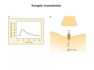
A hall mark of chemical transmission is a delay between presynaptic Ca2+ elevation and secretion. The delay can be as short as 0.2 ms, but is usually longer due to a variety of factors. Fundamental Neuro. 2002
Steps to chemical synaptic transmission. • First need to bring the presynaptic neuron to threshold at axon hillock. • Conduction down axon, length, R*C dependent. • Opening of voltage gated Ca channels. • Diffusion and action of Ca at release machinery. • Exocytosis and diffusion of transmitter in cleft. • Activation of postsynaptic receptors.
Synaptic delays can be less than 0.2 ms from calcium entry (Fund. Neurosci. Chap 8) to the beginning of secretion, but are typically longer when all steps (below) are considered. From Sudhof 2004
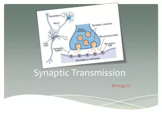
Chemical synapse types. • Axosomatic, axoaxonic, axodendritic, and dendrodendritic. • Excitatory (type I) and inhibitory (type II) synapses have different structure in CNS neurons. • CNS synapses usually have one or small number of release sites while nerve muscle synapses have up to 300 active zones. See http://synapses.mcg.edu/atlas
Type I Excitatory spherical Type II Inhibitory flattened See http://synapses.mcg.edu/atlas Josef Spacek
From Kristin Harris Lectures. http://synapses.mcg.edu/lab/harris/lectures.htm
Synapse structure like real estate location, location, location!! Multiple release sites NMJ From Squire et al. Fund. Neurosci 2nd ed.
Criteria for a chemical transmitter, make a case for glutamate. • The transmitter substance must be synthesized in the presynaptic neuron. Experiment? • It must be present in the presynaptic terminal and released in amounts sufficient to result in the level of response produced by the endogenous transmitter. experiment? • When applied exogenously the substance should mimic the effect of the endogenous transmitter. experiment? • A specific mechanism must exist for removing the transmitter from the synaptic cleft. experiment?
Ionic requirement for release. • Calcium influx is the trigger for fast evoked transmitter release • extracellular calcium (Ca2+) is typically about 1-2 mM, intracellular calcium at rest is <0.1 mM. • An elevation in intracellular calcium concentration is an absolute requirement for transmitter release. Na+ and K+ ions not necessary for release. • How do you test this hypothesis?
Ion substitution and pharmacology experiments. • The influx of calcium is triggered by voltage gated ion channels. • Depolarization itself is not needed to stimulate release, as calcium can be elevated by other means (caged calcium chelators). • The relationship between calcium influx and release is related to the "power (exponent)" of calcium entry and is highly nonlinear. For example for a 4th power relationship a doubling of calcium entry can produce a 16 fold increase in release.
Slope on a log-log plot indicates power relation, a small change in calcium produces a large change in release. Release~[Ca]^3-5 Slope=5.0 Slope=1.6 from Delaney Enc. of Sci.
Neurotransmitter release is triggered by a locally-activated low affinity sensor since bulk cytosolic [Ca2+]i rarely exceeds 10-6 M, yet transmitter release requires much higher [Ca2+]. G. Augustine Curr. Op. in Neurobiol.
Squid giant axon and release • Due to its large size the squid giant axon has been used to examine the calcium dependence of transmitter release. • Squid studies using different Ca2+ buffers indicate high concentrations of Ca2+ that are reached for less than 1 ms trigger release (Adler et al. 1991 J. Neurosci.). • Most calcium entry which triggers release occurs during the falling phase of action potential. Why is this advantageous?
Rate of calcium binding by buffer (chelator) provides insight into release machinery. • Fast BAPTA (kon 8x10⁸ M -1 *sec -1) buffers block release whereas, • slow EGTA (kon 1x10⁷ M-1*sec -1) buffers do not (Adler et al. 1991 J. Neurosci.). • Time for equilibration of EGTA with calcium~1000 ms versus 12.5 ms for BAPTA. • To estimate the buffer equilibration time take 1/(koff +kon*[Ca2+]), use koff of 8x101 sec –1 for both buffers and 1x10⁻⁴ for [Ca2+] at peak.
Temporal requirements. • Calcium trigger must be able to act within 0.1 ms of presynaptic stimulation. This requirement restricts the class of chemical events that may be involved in the evoked release process. • Phosphorylation, protein synthesis, gene expression all out. • Everything must be ready and localized- diffusion could not move transmitter vesicles or calcium very far.
Calcium channels are clustered on the release face (side with release) of the chick caylx synapse. Patch config. Outside of synapse Release face of synapse Reviewed in EF Stanley TINS 1997
Diffusion time of Ca2+ limits release latency. • Buffered calcium diffusion coeff. are on the order of 200 mm2/sec (D) so calcium could only diffuse a small distance at the most (~0.2 mm) during the synaptic delay (0.0001 s, 0.1 ms), so Ca2+ channels need to be very close to the release machinery. • distance=Sqrt(2Dt) D=diffusion coeff., t=time
The diffusion time is dependent on the square of the distance (d). • t=d2/(2*D), where t=time, d=distance, and D=diffusion coefficient. • For 0.1 mm the time is 0.025 ms. • However for 1.0 mm the time jumps by the square to 2.5 ms (100 times longer!), way longer than the release observed latency. These equations are for reference, the concept is of interest.
Kd or Km, concentration at ½ max binding or activity. Kd=Km=kdissoc/kassoc affinity=1/km, low affinity means a big km which usually means a large kdissoc Note koff=kdissoc and kon=kassoc
Low affinity binding gives rapid off rate. • Concentration for ½ max activity is ~1/affinity and is termed the Kd or Km, if a binding site has a low affinity more ligand is needed to get ½ saturation. • Therefore Kd=koff/kon, assuming a diffusion limited kon of 5x10⁸ M –1 s –1 then koff must be 5x10⁴ s-1 if the Kd is 100 mM. • To estimate the dissociation time constant take 1/(koff +kon*[Ca]) or 20 ms. Note after the channels close [Ca2+] is ~ 1x10⁻⁷ and the kon*[Ca] is small compared to koff. These equations are for reference.
For reference For a simple reaction. kon*[S] [E]+[S] [ES] koff The time constant for Ca2+ dissoc will be: t=1/(koff+kon*[S]) 20 ms=1/(5x104 s-1+ 5x10⁸ M –1 s –1 *1x10-7 M)
Local domains of Ca2+ near channel mouths control transmitter release. It is unclear whether release is always triggered by a single channel or whether multiple ones cooperate. EF Stanley TINS 1997
Transmitter release microdomains. Activation of Ca channels 1.8 mM outside Resting state just after channel closure, bulk Ca is up only 10%.
To make full use of microdomains the vesicle must be bound to the calcium channels. Atwood & Shanker Karunanithi DIVERSIFICATION OF SYNAPTIC STRENGTH: PRESYNAPTIC ELEMENTS Nature Reviews Neuroscience 3, 497 -516 (2002).
BAPTA, a faster buffer than EGTA more readily blocks synaptic transmission. Given 100 mM Ca2+, BAPTA equilibrates in <20 ms while EGTA takes ~1000 ms. B Atwood & Shanker Karunanithi DIVERSIFICATION OF SYNAPTIC STRENGTH: PRESYNAPTIC ELEMENTS Nature Reviews Neuroscience 3, 497 -516 (2002).
Calcium as a local messenger. • Fast channel activation, Ca2+ diffuses to couple excitation to synaptic chemistry. • Low affinity, rapid off rate, and restricted microdomains are characteristics of the calcium flux that evokes release. • Rapid off rate allows for re-loading of release mechanism when Ca2+ levels fall. Affinity=kon/koff • high off rate = low affinity.
Quantal aspects of release at the neuromuscular junction. • At the neuromuscular junction small spontaneous potentials (depolarizations) termed miniature end plate potentials are observed. Analogous to CNS miniature excitatory postsynaptic currents (mEPSCs). • The amplitude of evoked responses (due to calcium influx) is always an integer multiple of the unitary response. • Shown that calcium increases the probability of observing a unitary response and not its amplitude. • These data suggested the existence of transmitter quanta or packets. • Evoked transmission mediated by the release of ~ 150 quanta over a 1-3 ms period. Each quantum leads to about 0.5 mV depolarization.
From Kristin Harris Lectures. http://synapses.mcg.edu/lab/harris/lectures.htm
From Kristin Harris Lectures. http://synapses.mcg.edu/lab/harris/lectures.htm
From Kristin Harris Lectures. http://synapses.mcg.edu/lab/harris/lectures.htm
Stimulation Evoked amplitudes. 1X Miniature event histogram. 4X mini 2X 1X 3X 4X 2X 1 mV Squire Fund. Neurosci. 2002