Download
1 / 13
130 likes | 270 Views
Role of Estrogen Receptors in the Hippocampal-Neuropeptide Y Expression. Tamara Dennis Dr. Jana Velí š ková Albert Einstein College of Medicine. Introduction. Objective To determine the estrogen receptor (ER) type responsible for increasing the Neuropeptide Y (NPY) expression. Hypothesis

E N D
Role of Estrogen Receptors in the Hippocampal-Neuropeptide Y Expression. Tamara Dennis Dr. Jana Velíšková Albert Einstein College of Medicine
Introduction Objective • To determine the estrogen receptor (ER) type responsible for increasing the Neuropeptide Y (NPY) expression. Hypothesis • The ER is responsible for the expression of NPY.
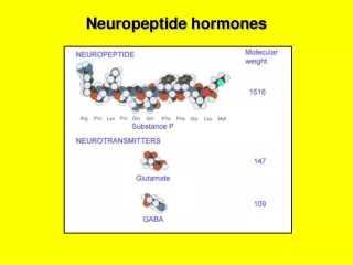
What are Estrogen Receptors? Estrogen receptors (ER) are intracellular proteins. The hormone estrogen binds to these receptors. There are two types of estrogen receptors, ER and ER . What is NPY? NPY is a powerful inhibitory peptide. It is located all over the brain. We are interested in the expression of NPY in the hippocampal hilar region. It is responsible for regulation of neuronal excitability, neuroprotection, feeding behaviors, circadian rhythm. Introduction
Role of NPY • It has been shown that during Status Epilepticus (SE) interneurons containing NPY are preferentially lost. • Interneurons are important inhibitory neurons. If they are lost, hyperexcitability and epileptogenesis occurs. • Increase of NPY in the hilus attenuates SE-induced damage to the hippocampus.
Methods & Procedures • Ovariectomy • Treat animals with DPN • Perfusion with formaldehyde & saline • Soak brains in formaldehyde overnight • Change solution to 30% sucrose • Immunohistochemistry • Cell counting under the microscope
DPN 2,3-bis(4-Hydroxyphenyl)-propionitrile ER agonist C15H13NO2 Drug
Histology Immunochemistry Day 1 50 l H2O2 + 5 ml PBS 30’ Wash 3x 5’ with PBS 200 l BSA + 200 l Triton + 500 l NGS + 4.228 ml PBS (double) 60’ 5 ml of solution 3 + 1.5 l of NPY antibody Day 2 Wash 3x 5’ with PBS 100 l BSA + 25 l anti-rabbit antibody + 4.9 ml PBS 60’ Prepare AB: 1drop of A and 1 drop of B in 5 ml PBS, Shake 30’ Wash 3x 5’ with PBS Apply AB solution 60’ Wash 3x with PBS 5’ DAB 8’ Immunohistochemistry
Results • Due to the time required to do this experiment there are no results as of yet.
Reibel S, Andre V, chassagnon S, Andre G, Marescaux C, Nehlig A, Depaulis A (200) Neuroprotective effects of chronic estradiol benzoate treatment on hippocampal cell loss induced by status epilepticus in the female rat. Neurosci Lett 281:79-82. Silva AP, Pinheiro PS, Carvalho AP, Carvalho CM, Jakobsen B, Zimmer J, Malva JO (2003) Activation of neuropiptide Y receptors is neuroprotective against excitotoxicity in organotypic hippocampal slice cultutes. FASEB J 17:1118- 1120. Sloviter RS (1994) The functional organization of the hippocampal dentate gyrus and its relevance to the pathogenesis of temporal lobe epilepsy. Ann Neurol 35:640-654. Sutula T, Cascino G, Cavazos J, Parada I, Ramirez L(1989) Mossy fiber synaptic reorganization in the epileptic human temporal lobe. Ann Neurol 26:321-330. Veliskova J (2003) The role of neuropiptide Y in estrogen-induced neuroprotective effects on status epilepticus-associated hippocampal cell loss. Epilepsia 44:206. Veliskova J, Velifsek L, Galanopoulou As, Sperber EF (2000) Neuroprotective effects od estrogens on hippocampal cells in adult female rats after statys epilepticus. Epilepsia 41:S30-S35. Vezzani A, Sperk G, Colmers WF (1999) neuropiptide Y:emerging evidence for a functional role in seizure modulation. Tends Neurosci 22:25-30. Ben-Ari, Tremblay E, Riche D, Ghilini G, Naquet R (1981) Electrographic, Clinical and pathological alterations following systematic administration of kainic acid, bicuclline or pentetrazole: metabolic mapping using the deoxyglucose method with special reference to the pathology of epilepsy. Neuroscience 6:1361-1391. Brandt C, Potschka H, Loscher W, Ebert U (2003) n-METHYL-d-aspartate receptor blockade after status epilepticus protects against limbic brain damage but not against epilepsy in the kainate model of temporal lobe epilepsy. Neuroscience 118:727-740. Buckmaster PS, Schwartzkroin PA (1995) interneurons and inhibition in the dentate gyrus of the rat in vivo. J Nuerosci 15:774-789. De Lanerolle NC, Kim JH, Robbins RJ, Spencer DD (1989) Hippocampus interneuron loss and plasticity in human temporal lobe epilepsy. Brain Res 495:387-395). El Bahh B, Auvergne R, Lere C, Brana C, Le Gal La Salle G, Rougier A (2001) Decreased epileptic susceptibility correlates with neuropiptide Y overexpression in a model of tolerance to excitotoxicity. Brain Res 894:209- 217. Freud TF, Buzsaki G (1996) Interneurons of the hippocampus. Hippocampus 6:347- 470. Galanopoulou AS, Alm EM, Veliskova J (2003) Estradiol reduces seixure-induced hippocampal injury in ovariectomized female but not in male rats. Neurosci Lett 342:201-205. References
Acknowledgements • Dr. Jana Velíšková • Dr. Sat Bhattacharya • Hong Wong • Albert Einstein College of Medicine • Harlem Children Society


![[IV] The Role of Chromatin Structure in Control of Gene Expression](https://cdn2.slideserve.com/4473274/slide1-dt.jpg)