Download
1 / 17
250 likes | 354 Views
Papulosquamous disorders. Pityriasis rosea lichen planus pityriasis rubra pilaris Erythroderma. Pityriasis rosea. may be caused by reactivation of either human herpes virus 7 or 6. The disease seems not to be contagious. It mainly affects children and young adults,

E N D
Papulosquamous disorders Pityriasisrosea lichen planus pityriasisrubrapilaris Erythroderma
Pityriasis rosea • may be caused by reactivation of either human herpes virus 7 or 6. • The disease seems not to be contagious. • It mainly affects children and young adults, • second attacks are rare. • About half of patients complain of itching. • A minority of patients have systemic symptoms such as aching and tiredness.
Most patients develop one plaque ( the ‘herald’ or ‘mother’ plaque) before the others. It is larger ( 2–5 cm diameter ) than later lesions, and is rounder, redder and more scaly.
After several days many smaller plaques appear, mainly on the trunk, but some also on the neck and extremities. An individual plaque is oval, salmon pink and shows a delicate scaling, adherent peripherally as a collarette.
The configuration of such plaques is characteristic. Their longitudinal axes run down and out from the spine in a ‘fir tree’ pattern , along the lines of the ribs.
eruption lasts 2–10 weeks and then resolves spontaneously • herald plaques are often mistaken for ringworm ( tineacorporis), the two disorders most likely to be misdiagnosed early in the general eruption are guttate psoriasis and secondary syphilis. • No treatment is curative. • A moderately potent topical steroid or calamine lotion will help the tching. Sunlight or artificial UVB often relieves pruritus and may hasten resolution. • treatment with antiviral agents has not been helpful.
lichen planus • Cause is unknown • Lichen planus is also associated with autoimmune disorders, such as alopecia areata, vitiligo and ulcerative colitis. • Contact allergy to mercury compounds ( in dental amalgam fillings) • Drugs too can cause lichen planus. • Some patients with lichen planus also have a hepatitis C infection .
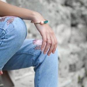
Typical lesions are violaceous or lilac-coloured, intensely itchy, flat-topped papules that usually arise on the extremities, particularly on the volar aspects of the wrists and legs . • A close look is needed to see a white streaky pattern on the surface of these papules ( Wickham’s striae).
White asymptomatic lacy lines, dots, and occasionally small white plaques, are also found in the mouth, particularly inside the cheeks, in about 50%of patients • oral lesions may be the sole manifestation of the disease.
patients rub rather than scratch, excoriations are uncommon. • Köbner phenomenon +ve. • The nails are usually normal, 10% of patients show changes ranging from fine longitudinal grooves to destruction of the entire nail fold and bed. • Scalp lesions can cause a patchy scarring alopecia.
the eruption as a whole tends to last about 1 year. • About one in six patients will have a recurrence. • Lichenoid drug reactions can mimic lichen planusclosely. antimalarials, beta-blockers, non-steroidal anti-inflammatory drugs, thiazide diuretics and penicillamine. • Contact with chemicals used to develop colour photographic film can also produce similar lesions. • The diagnosis is usually obvious clinically.
Potent topical steroids. • Systemic steroid in extensive involvement, nail destruction or painful erosive oral lichen planus. • photochemotherapywith psoralen and ultraviolet A ( PUVA ) or with narrowband UVB reduce pruritus and clear up the skin lesions. • Oral ciclosporin or acitretin • Antihistamines may blunt the itch. • Mucous membrane lesions, both oral and genital, applications of a corticosteroid or calcineurin inhibitor such as tacrolimus in a gel base
pityriasis rubra pilaris • uncommon skin disorders characterized by fine scaling (pityriasis), redness (rubra) and involvement of hair follicles ( pilaris). • No cause has been identified. • familial type: autosomal dominant inheritance.develops gradually in childhood. • The most common acquired form generally resolves within 3 years, but may recur. • The familial type, developing in childhood, persists throughout life.
erythema and scaling of the face and scalp. Later, red or orange plaques grow quickly and merge, so that patients with pityriasisrubrapilaris are often erythrodermic. • Perifollicular papules keratinous follicular plugs • Small islands of skin may be ‘spared’ • palms and soles become thickened, smooth and yellow.
Emollient and keratolytics forpalms and soles . • About 50% of patients respond slowly to systemic retinoids such as acitretin ( in adults, 25–50 mg/day for 6–8 months). • Oral methotrexate in low doses, taken once a week. • Phototherapiesdo not help much. • Systemic steroids are not indicated.
Erythroderma • ‘Erythroderma’ is the term used when the skin is red with little or no scaling, while the term ‘exfoliative dermatitis’ is preferred if scaling predominates. • If chronic, tightness of the facial skin leadsto ectropion, scalp and body hair may be lost, nails may be shed too. • Temperature regulation is impaired , patient feel cold and shiver. Oedema, tachycardia, anaemia, dehydration can occur.
Some causes of erythroderma/exfoliativedermatitis • Psoriasis • Pityriasisrubrapilaris • Ichthyosiformerythroderma • Pemphiguserythematosus • Contact, atopic or seborrhoeic eczema • Drug eruptions • Crusted (Norwegian) scabies