Download
1 / 12
120 likes | 385 Views
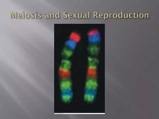
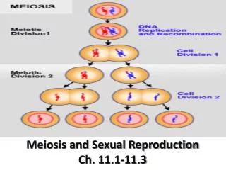
Chapter 13: Meiosis and Sexual Life Cycles. Parent. Bud. 0.5 mm. Figure 13.2 The asexual reproduction of a hydra. APPLICATION A karyotype is a display of condensed chromosomes arranged in pairs. Karyotyping can be used to screen for abnormal numbers of chromosomes or

E N D
Chapter 13: Meiosis and Sexual Life Cycles
Parent Bud 0.5 mm Figure 13.2 The asexual reproduction of a hydra
APPLICATION A karyotype is a display of condensed chromosomes arranged in pairs. Karyotyping can be used to screen for abnormal numbers of chromosomes or defective chromosomes associated with certain congenital disorders, such as Down syndrome. TECHNIQUE Karyotypes are prepared from isolated somatic cells, which are treated with a drug to stimulate mitosis and then grown in culture for several days. A slide of cells arrested in metaphase is stained and then viewed with a microscope equipped with a digital camera. A digital photograph of the chromosomes is entered into a computer, and the chromosomes are electronically rearranged into pairs according to size and shape. Pair of homologous chromosomes 5 µm Centromere RESULTS This karyotype shows the chromosomes from a normal human male. The patterns of stained bands help identify specific chromosomes and parts of chromosomes. Although difficult to discern in the karyotype, each metaphase chromosome consists of two, closely attached sister chromatids (see diagram). Sister chromatids Figure 13.3 Preparing a Karyotype
Key Maternal set of chromosomes (n = 3) 2n = 6 Paternal set of chromosomes (n = 3) Two sister chromatids of one replicated chromosome Centromere Two nonsister chromatids in a homologous pair Pair of homologous chromosomes (one from each set) Figure 13.4 Describing chromosomes
Key Haploid gametes (n = 23) Haploid (n) Ovum (n) Diploid (2n) Sperm Cell (n) FERTILIZATION MEIOSIS Ovary Diploid zygote (2n = 46) Testis Mitosis and development Multicellular diploid adults (2n = 46) Figure 13.5 The human life cycle
Key Haploid Diploid Haploid multicellular organism Haploid multicellular organism (gametophyte) Gametes n n n Mitosis Mitosis Mitosis Mitosis n n n n n n n n MEIOSIS FERTILIZATION n Spores Gametes n Gametes MEIOSIS FERTILIZATION MEIOSIS FERTILIZATION Zygote 2n 2n 2n 2n Diploid multicellular organism (sporophyte) Zygote 2n Diploid multicellular organism Mitosis Mitosis Zygote (b) Plants and some algae (a) Animals (c) Most fungi and some protists Figure 13.6 Three types of sexual life cycles
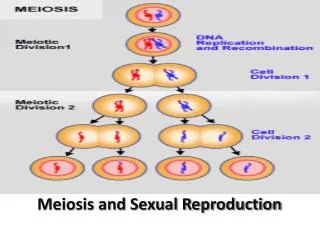
Interphase Homologous pair of chromosomes in diploid parent cell Chromosomes replicate Homologous pair of replicated chromosomes Sister chromatids Diploid cell with replicated chromosomes Meiosis I 1 Homologous chromosomes separate Haploid cells with replicated chromosomes Meiosis II 2 Sister chromatids separate Haploid cells with unreplicated chromosomes Figure 13.7 Overview of meiosis: how meiosis reduces chromosome number
MEIOSIS I: Separates homologous chromosomes INTERPHASE PROPHASE I METAPHASE I ANAPHASE I Sister chromatids remain attached Centromere (with kinetochore) Centrosomes (with centriole pairs) Chiasmata Metaphase plate Sister chromatids Spindle Nuclear envelope Homologous chromosomes separate Microtubule attached to kinetochore Tetrad Chromatin Tetrads line up Homologous chromosomes (red and blue) pair and exchange segments; 2n = 6 in this example Pairs of homologous chromosomes split up Chromosomes duplicate Figure 13.8 The Meiotic Division of an Animal Cell
MEIOSIS II: Separates sister chromatids TELOPHASE II AND CYTOKINESIS TELOPHASE I AND CYTOKINESIS METAPHASE II ANAPHASE II PROPHASE II Cleavage furrow Haploid daughter cells forming Sister chromatids separate Two haploid cells form; chromosomes are still double During another round of cell division, the sister chromatids finally separate; four haploid daughter cells result, containing single chromosomes Figure 13.8 The Meiotic Division of an Animal Cell
MITOSIS MEIOSIS Chiasma (site of crossing over) Parent cell (before chromosome replication) MEIOSIS I Prophase I Prophase Chromosome replication Chromosome replication Tetrad formed by synapsis of homologous chromosomes Duplicated chromosome (two sister chromatids) 2n = 6 Tetrads positioned at the metaphase plate Chromosomes positioned at the metaphase plate Metaphase I Metaphase Sister chromatids separate during anaphase Anaphase Telophase Homologues separate during anaphase I; sister chromatids remain together Anaphase I Telophase I Haploid n = 3 Daughter cells of meiosis I 2n 2n MEIOSIS II Daughter cells of mitosis n n n n Daughter cells of meiosis II Sister chromatids separate during anaphase II Figure 13.9 A comparison of mitosis and meiosis
Key Maternal set of chromosomes Possibility 1 Possibility 2 Paternal set of chromosomes Two equally probable arrangements of chromosomes at metaphase I Metaphase II Daughter cells Combination 1 Combination 2 Combination 3 Combination 4 Figure 13.10 The independent assortment of homologous chromosomes in meiosis
Prophase I of meiosis Nonsister chromatids Tetrad Chiasma, site of crossing over Metaphase I Metaphase II Daughter cells Recombinant chromosomes Figure 13.11 The results of crossing over during meiosis