Download
1 / 24
1.02k likes | 3.16k Views
Inguinal Canal. Prepared by: Dr. Azmi hussien PhD. Md. Head of human anatomy Department. Inguinal Canal. The inguinal canal is an oblique passage through the lower part of the anterior abdominal wall. In the males, it allows structures to pass to and from the testis to the abdomen.

E N D
Inguinal Canal Prepared by: Dr. Azmihussien PhD. Md. Head of human anatomy Department
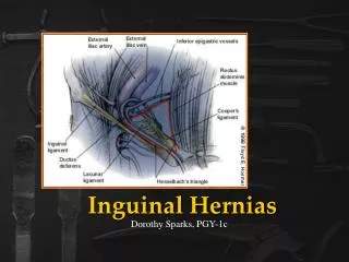
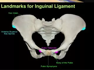
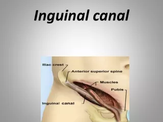
Inguinal Canal • The inguinal canal is an oblique passage through the lower part of the anterior abdominal wall. • In the males, it allows structures to pass to and from the testis to the abdomen. • females it allows the round ligament of the uterus to pass from the uterus to the labium majus. • The canal is about 1.5 in. (4 cm) long in the adult and extends from the deep inguinal ring, a hole in the fascia transversalis, downward and medially to the superficial inguinal ring, a hole in the aponeurosis of the external oblique muscle.
The deep inguinal ring: an oval opening in the fascia transversalis, lies about 0.5 in. (1.3 cm) above the inguinal ligament midway between the anterior superior iliac spine and the symphysis pubis. • Related to it medially are the inferior epigastric vessels, which pass upward from the external iliac vessels. • The margins of the ring give attachment to the internal spermatic fascia (or the internal covering of the round ligament of the uterus). • The superficial inguinal ring: is a triangular-shaped defect in the aponeurosis of the external oblique muscle and lies immediately above and medial to the pubic tubercle. • The margins of the ring, sometimes called the crura, give attachment to the external spermatic fascia.
Walls of the Inguinal Canal • Anterior wall: External oblique aponeurosis, reinforced laterally by the origin of the internal oblique from the inguinal ligament .. • Posterior wall: Conjoint tendon medially, fascia transversalis laterally . • Roof or superior wall: Arching lowest fibers of the internal oblique and transversusabdominis muscles. • Floor or inferior wall: Upturned lower edge of the inguinal ligament and, at its medial end, the lacunar ligament.
Spermatic Cord • The spermatic cord is a collection of structures that pass through the inguinal canal to and from the testis. • It begins at the deep inguinal ring lateral to the inferior epigastric artery and ends at the testis.
Structures of the Spermatic Cord The structures are as follows: • Vas deferens • Testicular artery • Testicular veins (pampiniform plexus) • Testicular lymph vessels • Autonomic nerves • Remains of the processusvaginalis • Genital branch of the genitofemoral nerve, which supplies the cremaster muscle
Scrotum, Testis, and Epididymides • Scrotum • The scrotum is an outpouching of the lower part of the anterior abdominal wall. • It contains the testis, the epididymides, and the lower ends of the spermatic cords.
The wall of the scrotum has the following layers: • Skin. • Superficial fascia: This is continuous with the fatty and membranous layers of the anterior abdominal wall. • the fat is replaced by smooth muscle called the dartos muscle. • This is innervated by sympathetic nerve fibers and is responsible for the wrinkling of the overlying skin. • The membranous layer of the superficial fascia (often referred to as Colles' fascia) is continuous in front with the membranous layer of the anterior abdominal wall (Scarpa's fascia),
3. Spermatic fasciae: These three layers lie beneath the superficial fascia and are derived from the three layers of the anterior abdominal wall on each side, as previously explained.
The external spermatic fascia is derived from the aponeurosis of the external oblique muscle; • the cremasteric fascia is derived from the internal oblique muscle • the internal spermatic fascia is derived from the fascia transversalis. The cremaster muscle is supplied by the genital branch of the genitofemoral nerve. • The cremaster muscle can be made to contract by stroking the skin on the medial aspect of the thigh. This is called the cremasteric reflex
5-Tunica vaginalis: This lies within the spermatic fasciae and covers the anterior, medial, and lateral surfaces of each testis.
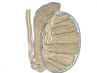
Testis • The testis is a firm, mobile organ lying within the scrotum. • The left testis usually lies at a lower level than the right. • Each testis is surrounded by a tough fibrous capsule, the tunica albuginea. • Extending from the inner surface of the capsule is a series of fibrous septa that divide the interior of the organ into lobules. • Lying within each lobule are one to three coiled seminiferous tubules. • The tubules open into a network of channels called the rete testis. Small efferent ductulesconnect the rete testis to the upper end of the epididymis .
Epididymis • The epididymis is a firm structure lying posterior to the testis, with the vas deferens lying on its medial side. • It has an expanded upper end, the head, a body, and a pointed tail inferiorly. • Laterally, a distinct groove lies between the testis and the epididymis, which is lined with the inner visceral layer of the tunica vaginalis and is called the sinus of the epididymis.
The epididymis is a much coiled tube nearly 20 ft (6 m) long, embedded in connective tissue. • The tube emerges from the tail of the epididymis as the vas deferens, which enters the spermatic cord. • The long length of the duct of the epididymis provides storage space for the spermatozoa and allows them to mature. • A main function of the epididymis is the absorption of fluid. addition of substances to the seminal fluid to nourish the maturing sperm.
Blood Supply of the Testis and Epididymis • The testicular artery is a branch of the abdominal aorta. • The testicular veins emerge from the testis and the epididymis as a venous network, the pampiniform plexus. • This becomes reduced to a single vein as it ascends through the inguinal canal. • The right testicular vein drains into the inferior vena cava, and the left vein joins the left renal vein.