Download
1 / 32
330 likes | 558 Views
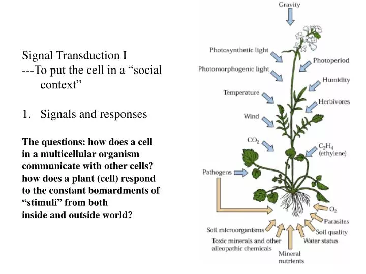
Signal Transduction I ---To put the cell in a “social context” Signals and responses The questions: how does a cell in a multicellular organism communicate with other cells? how does a plant (cell) respond to the constant bomardments of “stimuli” from both inside and outside world?.

E N D
Signal Transduction I • ---To put the cell in a “social context” • Signals and responses • The questions: how does a cell • in a multicellular organism • communicate with other cells? • how does a plant (cell) respond • to the constant bomardments of • “stimuli” from both • inside and outside world?
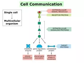
2) The language: chemical, electrical, physical contact 3) Partners: cell-cell, cell-environment, plant-other organisms Signaling cell target cell 4) The receptors and specificity cell surface vs intracellular, specificity of signaling
5) The pathways and networks: molecular relays are weaved into a “network” or Circuit in the Communication Processes: The processes to bring changes into the system. The changes will Be integrated to the “response” as a way to adapt to the signals… Responses
2. Major pathways in eukaryotes as defined by surface receptors 1) The G- protein coupled receptors 2) The enzyme receptors 3) Ion channel As receptors
The G-protein coupled receptors • A number of hormones or neurotransmittors utilize this type of the receptors • The receptor is a • seven-transmembrane protein • c) Binding of ligand to the receptor • Triggers Interaction • With G protein • d) The trimeric • G protein consists • Of alpha, beta and • Gamma subunits • e) Active state is the • GTP-binding form and • Inactive state is the GDP form. • f) GTP binding dissociates the 3 subunits • And activate the G protein. GTP • Hydrolysis into GDP inactivates it.
2) HOW TO STUDY RECEPTORS? • Binding assay: to see where is the receptor, one can utilize a labeled ligand to do the binding assay—you can fractionate the cells to purify specific organelle of membrane to mix with the ligand and see if it binds to the fraction. Three parameters will be critical: the specificity and affinity should be consistent with the biological function of the ligand. The binding should be “saturable” because there is only a limited number of the receptor in a fraction. Then purify the receptor protein and clone the genes. total specific amount Non-specific (cannot be competed by the unlabeled ligand) Concentration (biological?)
B) Functional cloning of the receptor by randomly expressing a cDNA library in a model system (eg, oocytes or culture cells): monitor the function of the receptor by adding the ligand and measure specific response in the expression system. Retrieve the specific mRNA that causes the response
3) The targets for trimeric G proteins and production of second messengers • The cAMP pathway: • The beta-adrenergic receptor binds adrenaline and activates a G protein that in turn activates adenylyl cyclase that forms cAMP from ATP, cAMP functions as second messenger to activate a protein kinase called PKA, PKA phosphorylates glycogen phosphorylase kinase (GPK), this kinase then phosphorylates glycogen phosphorylase (GP) that degrades glycogen into glucose---blood sugar level increases—the response. [glucose] glycogen GP ATPcAMPPKAGPK
How does cAMP work to activate PKA? What is a second messenger? cAMP C R C R C R active C R Inactive C R C R 2nd messenger: small molecule that is produced by the primary signal and functions to pass the signal further downstream to cellular response. b) The IP3-Ca2+ pathway: The trimeric G proteins are present in many forms (different proteins encoded by different genes). Some forms target adenylyl cyclase, and some other forms target other enzymes such as PLC (phospholipase C), PLC cleaves phosphotidylinositol 4,5 triphosphate (PIP2) and produce IP3 and Diacylglycerol (DAG), these second messengers activate different targets---IP3 activates a calcium channel in the ER membrane and release calcium into the cytosol and DAG activates a protein kinase called PKC. Calcium can be sensed by protein sensors such as calmodulin (CaM) that binds calcium and change conformation, CaM then activate many other proteins including a protein kinase that phosphorylates myosin light chain and activate the muscle activity.
Calcium is a critical Second messenger in Plant cells. It is very Tightly regulated by Many proteins Like channels and pumps Many organelles Are involved in Calcium homeo- stasis
Measuring calcium levels in living cells: fluorescent dye, aequorin, cameolin (CaM-GFP), etc Pollen tube growth and calcium wave
How does calcium works? Calcium sensors detect the changes in calcium concentration by Binding it and conformational changes. Such change will trigger Interaction with downstream targets such as protein kinases that modify other protein activity. CaM takes a new structure when in calcium-bound form. A specific sequence in CaM forms the Ca-binding “hand” shown at right.
DAG—the other second messenger produced from PLC activity The target PKC needs calcium in order to get associated with the Membrane where DAG activates the kinase activity
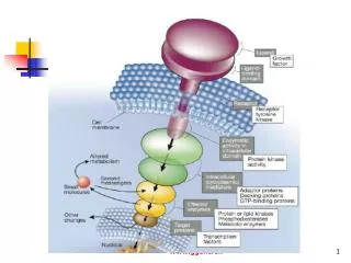
2) The enzyme receptors— receptor kinases as example Many growth factors in animals such as epidermal growth factor (EGF) work by binding to their surface receptors that are tyrosine kinases. Binding triggers dimerization of the receptor and activation of the kinase activity. The kinases phosphorylate each other and produce a “highly phosphosphorlyated cluster” that serves as a “module” for “specific interaction” with the adaptor proteins. These proteins then interact with other proteins and pass the signal downstream to a small G protein called Ras, one of the earliest oncogenes to be discovered. Ras activates Raf, a ser/thr kinase that activate MEK, another kinase, that activates MAPK, still another kinase. MAPK activates proteins that activate gene expression---response. See the picture next page.
How phosphorylation changes MAPK structure (spot the major difference of these two structures)
How does MAPK cascade determine the specificity of signals? --The scaffold proteins hold the key • Different isoforms in each step of the relay • Different scaffold proteins recruit • Different combination of the relay molecules • c) Different signal and outcome
3) RLKs in plants---a large family of receptor proteins (<700) RLKs (Receptor-Like Kinases): contains an extracellular domain, a transmembrane domain, and a cytoplasmic kinase domain It is hypothesized that RLKs work Like tyrosine kinase receptors in Animals and form dimer after ligand Binding. Kinase becomes activated and phosphorylates each other to form A binding module to recruit other Proteins onto the membrane. It is also believed that MAPK cascade Is downstream from RLK.
The apical meristem development involves a RLK pathway • Genetics analysis of meristem size identified mutants that produce larger meristems and more flowers. One group of mutants called clavata 1 are mutated in a RLK gene, clavata 2 is mutated in a RLK-like gene (everything like RLK but without the kinase domain), the third mutant is mutated in a gene encoding a small protein localized to the cell wall. Further study showed that CLV1/CLV2/CLV3 are present in a supermolecular complex in the plasma membrane. This complex also recruits other proteins including a protein phosphatase (KAPP), and a small G protein Rop. Rop may be like Ras that pass the RLK signal down to MAPK cascade.
WT clv1 Second example is stomatal development that appears to be controlled by RLK as well. Third example is the symbiotic interaction: RLK serves as receptor for a nodulation factor from bacterial cells. Fourth, you already heard about WAKs and their role in cell expansion—they belong to RLK super-family also. The list will go on and on as more work is done with the functional analysis of the RLK genes. Friday lecture will give you at least one more example…
4). From cell surface to the nucleus cAMP pathway and receptor kinase pathway not only would modify the cellular components in the cytoplasm but in many cases they can lead the signaling processes into the nucleus to modify gene expression. PKA catalytic Subunit (c) move into the nucleus and phospholrylates the transcription factor CREB that forms protein complex with P300 and turns on gene transcription
Similarly, the MAPK can be transported into the nucleus after phosphorylated by MAPKK. In nucleus it also phosphorylate some transcriptional proteins for gene activation.
5) Signals must be removed after activation --Cell signaling is defined by temporal and spatial information. It is not always on but rather have “frequency”—on and off, on again and off again… To keep a frequency, “off” is equally important—the signal must be removed. --Very often the signal is not present but arrives when needed---so when the signal is not there anymore, the pathway will be shut down. This shutting down process also involves “removal” of active molecules. Simplest and could be most important example: kinase and phosphatase work in “pairs” to modify the protein structure/function by putting on or taking off the phosphate from the protein. Another one is the calcium: elevation in the cytosol and removed by sequestering it into the vacuole or other organelles. Third one is the G protein switch: GTP vs GDP binding forms
3. Crosstalk and signaling networks Pathways in the cell interact with each other and regulate each other. Therefore, the “linear” pathways are weaved into a complex network like a public transportation network that consists of buses, subways, and trains… A component in the network can be considered as a “knot” on the network (a fishnet). All components are connected to each other directly or indirectly. Anything wrong with one component may affect the “status” of other components.
An example of pathway Interaction: Protein kinases in the G- Protein coupled receptor Pathway can regulate the Activity of MAPK and Vise versa. The kinases could also Share a common protein As substrate so the path- Way may converge at this Protein substrate.
4. Evolution of signaling pathways: from bacteria to human • The two component system dominates the bacterial world: • The signal receptor and response regulator---phosphate transfer from histidine to aspartate 2) This system was passed on to plants and fungi but not animals The histidine kinase and the response regulators have “fused” together in many cases although some separate response regulators still exist in fungi and plants.
3) Many new paradigms appeared in eukaryotic systems Think about the complexity of the cell structure…from prokaryotes to eukaryotes The G-protein coupled receptors dominate the animal signaling, but very few pathways in fungi and plants use this. The tyrosine kinase receptors have never been found in fungi and plants! Plants developed a large number of ser/thr receptor kinases that are not a major theme in animals.