Download
1 / 60
600 likes | 607 Views
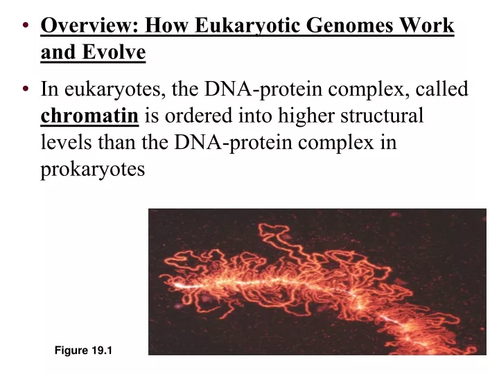
Overview: How Eukaryotic Genomes Work and Evolve In eukaryotes, the DNA-protein complex, called chromatin is ordered into higher structural levels than the DNA-protein complex in prokaryotes. Figure 19.1. Chromatin in a Developing Salamander Ovum. Chromatin, detail.

E N D
Overview: How Eukaryotic Genomes Work and Evolve In eukaryotes, the DNA-protein complex, called chromatin is ordered into higher structural levels than the DNA-protein complex in prokaryotes Figure 19.1
Gene Regulation Resources: Gene Switches from HHMI: http://www.hhmi.org/biointeractive/gene-switch Epigenetics from Genetic Science Learning Center: http://learn.genetics.utah.edu/content/epigenetics/ Epigenetics on PBS Including NOVA Science NOW: http://www.pbs.org/wgbh/nova/body/epigenetics.html Epigenetics from Bozeman Science (Paul Andersen): http://www.bozemanscience.com/epigenetics/?rq=epigenetics Genetic Switches Click and Learn from HHMI: http://www.hhmi.org/biointeractive/genetic-switches RNAi from PBS (NOVA) http://www.pbs.org/wgbh/nova/body/rnai.html RNAi Animation - McGraw Hill https://highered.mheducation.com/sites/9834092339/student_view0/chapter16/rna_interference.html RNAi on YouTube: https://www.youtube.com/watch?v=2dL7Sh_udKs&feature=youtu.be RNAin on Nature: https://www.youtube.com/watch?v=cK-OGB1_ELE Transposan Animation - McGraw Hill http://highered.mheducation.com/sites/0072556781/student_view0/chapter13/animation_quiz_5.html Transcription Complex and Enhancers: https://highered.mheducation.com/sites/9834092339/student_view0/chapter16/transcription_complex_and_enhancers.html DNA Packing Animation from HHMI: http://www.hhmi.org/biointeractive/dna-packaging DNA Packing from Dolan DNA Learning Center: https://www.dnalc.org/view/15483-DNA-packaging-3D-animation-with-basic-narration.html Proteasome Action from HHMI:http://www.hhmi.org/biointeractive/proteasome Marine vs. Freshwater Sticklebacks - HHMI Animation https://www.youtube.com/watch?v=p0jEkOT-eB0 X-Inactivation Animation: http://www.hhmi.org/biointeractive/x-inactivation
Both prokaryotes and eukaryotes Must alter their patterns of gene expression in response to changes in environmental conditions
Concept 19.1: Chromatin structure is based on successive levels of DNA packing Eukaryotic DNA Is precisely combined with a large amount of protein Eukaryotic chromosomes Contain an enormous amount of DNA relative to their condensed length
Proteins called histonesare responsible for the first level of DNA packing in chromatin Bind tightly to DNA The association of DNA and histones seems to remain intact throughout the cell cycle Nucleosomes, or “Beads on a String”
Degree of packing of DNA regulates transcription tightly wrapped around histones no transcription genes turned off DNA Packing as Gene Control • heterochromatin darker DNA (H) = tightly packed • euchromatin lighter DNA (E) = loosely packed H E
In electron micrographs Unfolded chromatin has the appearance of beads on a string Each “bead” is a nucleosome, the basic unit of DNA packing 2 nm DNA double helix Histone tails His- tones 10 nm Histone H1 Linker DNA (“string”) Nucleosome (“bad”) (a) Nucleosomes (10-nm fiber) Figure 19.2 a
“Beads on a string” first level of DNA packing histone proteins 8 protein molecules many positively charged amino acids bind tightly to negatively charged DNA 8 histone molecules Nucleosomes
Polar, Acidic, and Basic Amino Acids Generally positive in charge Generally negative in charge
Concept 19.2: Gene expression can be regulated at any stage, but the key step is transcription All organisms must regulate which genes are expressed at any given time During development of a multicellular organism cells undergo a process of specialization in form and function called cellular differentiation
Each cell of a multicellular eukaryote expresses only a fraction of its genes In each type of differentiated cell a unique subset of genes is expressed to make the 200 different cell types in a human Differential Gene Expression
Many key stages of gene expressionCan be regulated in eukaryotic cells
Chemical modification of histone tails can affect the configuration of chromatin and thus gene expression Chromatin changes Transcription RNA processing Translation mRNA degradation Protein processing and degradation Histone tails DNA double helix Amino acids available for chemical modification Histone Modification Figure 19.4a (a) Histone tails protrude outward from a nucleosome
Histone Acetylation Seems to loosen chromatin structure and thereby enhance transcription Acetylated histones Unacetylated histones (b) Acetylation of histone tails promotes loose chromatin structure that permits transcription Figure 19.4 b
Histone Acetylation • Acetylation of histones unwinds DNA • loosely wrapped around histones • enables transcription • genes turned on • attachment of acetyl groups (–COCH3) to histones • conformational change in histone proteins • transcription factors have easier access to genes
Addition of methyl groups to certain bases in DNA is associated with reduced transcription in some species DNA Methylation
Methylation of DNA blocks transcription factors no transcription genes turned off attachment of methyl groups (–CH3) to cytosine nearly permanent inactivation of genes ex. inactivated mammalian X chromosome = Barr body DNA Methylation
EpigeneticInheritance is the inheritance of traits transmitted by mechanisms not directly involving the nucleotide sequence
Associated with most eukaryotic genes are multiple control elements Segments of noncoding DNA that help regulate transcription by binding certain proteins Poly-A signal sequence Termination region Proximal control elements Enhancer (distal control elements) Exon Intron Intron Exon Exon DNA Downstream Upstream Promoter Transcription Poly-A signal Exon Exon Intron Intron Exon Cleared 3 end of primary transport Primary RNA transcript (pre-mRNA) 5 Chromatin changes RNA processing: Cap and tail added; introns excised and exons spliced together Transcription Intron RNA RNA processing Coding segment mRNA degradation Translation mRNA P Protein processing and degradation G P P Start codon Poly-A tail Stop codon 3 UTR (untranslated region) 5 Cap 5 UTR (untranslated region) Figure 19.5
Proximal control elements are located close to the promoter Distal control elements, groups of which are called enhancers may be far away from a gene or even in an intron
Transcription Complex Activator Proteins • regulatory proteins bind to DNA at distant enhancer sites • increase the rate of transcription Enhancer Sites regulatory sites on DNA distant from gene Enhancer Activator Activator Activator Coactivator E F B RNA polymerase II H TFIID A Coding region T A T A Basal Transcription Factor Core promoter and initiation complex Initiation Complex at Promoter Site binding site of RNA polymerase
Enhancer DNA sequences distant control sequences Activator proteins bind to enhancer sequence & stimulates transcription Silencer proteins bind to enhancer sequence & block gene transcription Model for Enhancer Action
Activators are proteins that bind to enhancers and stimulate transcription of a gene Distal control element Promoter Activators Gene Enhancer TATA box General transcription factors Activator proteins bind to distal control elements grouped as an enhancer in the DNA. This enhancer has three binding sites. 1 DNA-bending protein Group of Mediator proteins A DNA-bending protein brings the bound activators closer to the promoter. Other transcription factors, mediator proteins, and RNA polymerase are nearby. 2 RNA Polymerase II Chromatin changes The activators bind to certain general transcription factors and mediator proteins, helping them form an active transcription initiation complex on the promoter. 3 Transcription RNA processing RNA Polymerase II mRNA degradation Translation Protein processing and degradation Transcription Initiation complex RNA synthesis Figure 19.6
Some specific transcription factors function as repressors to inhibit expression of a particular gene Some activators and repressors act indirectly by influencing chromatin structure
Alternative RNA splicing variable processing of exons creates a family of proteins, depending on which RNA segments are treated as exons and which as introns Post-Transcriptional Control
MicroRNAs (miRNAs) • small single-stranded RNA molecules that can bind to mRNA • These can degrade mRNA or block its translation • Inhibition of gene expression by RNA molecules = RNA INTERFERENCE (RNAi)
RNA interference by single-stranded microRNAs (miRNAs) Can lead to degradation of an mRNA or block its translation The bound miRNA can base-pair with any target mRNA that contains the complementary sequence. The micro- RNA (miRNA) precursor folds back on itself, held together by hydrogen bonds. One strand of each short double- stranded RNA is degraded; the other strand (miRNA) then associates with a complex of proteins. 3 The miRNA-protein complex prevents gene expression either by degrading the target mRNA or by blocking its translation. An enzyme called Dicer moves along the double- stranded RNA, cutting it into shorter segments. 4 2 1 5 2 Chromatin changes Transcription RNA processing mRNA degradation Translation Protein complex Protein processing and degradation Dicer Degradation of mRNA OR miRNA Target mRNA Blockage of translation Hydrogen bond 5 Figure 19.9
Figure 18.15 Hairpin Hydrogenbond miRNA Dicer 5 3 (a) Primary miRNA transcript miRNA miRNA-proteincomplex mRNA degraded Translation blocked (b) Generation and function of miRNAs
Small Interfering RNAs (siRNAs) • RNA interference (RNAi) is caused by siRNAs • Ex: Yeast: siRNA’s play a role in heterochromatin formation and can block large regions of the chromosome
The initiation of translation of selected mRNAs can be blocked by regulatory proteins that bind to specific sequences or structures of the mRNA Alternatively, translation of all the mRNAs in a cell may be regulated simultaneously
After translation various types of protein processing, including cleavage and the addition of chemical groups, are subject to control
“Death tag” mark unwanted proteins with a label 76 amino acid polypeptide, ubiquitin labeled proteins are broken down rapidly in "waste disposers" proteasomes Ubiquitin Aaron Ciechanover Israel Avram Hershko Israel Irwin Rose UC Riverside
Proteasomes Are giant protein complexes that bind protein molecules and degrade them Enzymatic components of the proteasome cut the protein into small peptides, which can be further degraded by other enzymes in the cytosol. 3 The ubiquitin-tagged protein is recognized by a proteasome, which unfolds the protein and sequesters it within a central cavity. 2 1 Multiple ubiquitin mol- ecules are attached to a protein by enzymes in the cytosol. Chromatin changes Transcription RNA processing Proteasome and ubiquitin to be recycled Ubiquitin Translation mRNA degradation Proteasome Protein processing and degradation Protein fragments (peptides) Protein to be degraded Ubiquinated protein Protein entering a proteasome Figure 19.10
Protein-degrading “machine” cell’s waste disposer breaks down any proteins into 7-9 amino acid fragments cellular recycling Proteasome
Concept 19.4 Eukaryotic genomes can have many noncoding DNA sequences in addition to genes The bulk of most eukaryotic genomes consists of noncoding DNA sequences, often described in the past as “junk DNA” However, much evidence is accumulating that noncoding DNA plays important roles in the cell
Compared with prokaryotic genomes, the genomes of eukaryotes Generally are larger Have longer genes Contain a much greater amount of noncoding DNA both associated with genes and between genes The Relationship Between Genomic Composition and Organismal Complexity
Now that the complete sequence of the human genome is available We know what makes up most of the 97-98% that does not code for proteins, rRNAs, or tRNAs Exons (regions of genes coding for protein, rRNA, tRNA) (1.5%) Repetitive DNA that includes transposable elements and related sequences (44%) Introns and regulatory sequences (24%) Unique noncoding DNA (15%) Repetitive DNA unrelated to transposable elements (about 15%) Alu elements (10%) Simple sequence DNA (3%) Large-segment duplications (5-6%) Figure 19.14
The first evidence for wandering DNA segments Came from geneticist Barbara McClintock’s breeding experiments with Indian corn Figure 19.15
Eukaryotic transposable elements are of two types: Transposons, which move within a genome by means of a DNA intermediate Retrotransposons, which move by means of an RNA intermediate New copy of transposon Transposon DNA of genome Transposon is copied Insertion Mobile transposon (a) Transposon movement (“copy-and-paste” mechanism) New copy of retrotransposon Retrotransposon DNA of genome RNA Insertion Reverse transcriptase (b) Retrotransposon movement Figure 19.16a, b
A particular exon within a gene could be duplicated on one chromosome and deleted from the homologous chromosome Rearrangements of Parts of Genes: Exon Duplication and Exon Shuffling
In exon shuffling errors in meiotic recombination lead to the occasional mixing and matching of different exons either within a gene or between two nonallelic genes EGF EGF EGF EGF Epidermal growth factor gene with multiple EGF exons (green) Exon shuffling Exon duplication F F F F Fibronectin gene with multiple “finger” exons (orange) F EGF K K K Exon shuffling Plasminogen gene with a “kfingle” exon (blue) Portions of ancestral genes TPA gene as it exists today Figure 19.20