Download
1 / 38
390 likes | 539 Views
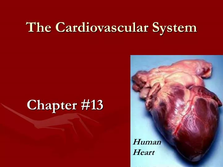
The Cardiovascular System. Chapter #13. Human Heart. 13.1 Introduction. The heart pump 7,000 liters of blood/day. Pumps 2.5 billion times in an average life span. The heart, arteries, veins, capillaries, and blood make up the cardiovascular system.

E N D
The Cardiovascular System Chapter #13 Human Heart
13.1 Introduction • The heart pump 7,000 liters of blood/day. • Pumps 2.5 billion times in an average life span. • The heart, arteries, veins, capillaries, and blood make up the cardiovascular system. • The main job of the cardiovascular system is to deliver oxygen and nutrients to all body cells and remove waste from all cells. • Without fresh O2 and removing waste death would be quick.
13.2 Structure of the Heart • The heart is a hollow, cone-shaped, muscular pump located within the thoracic cavity and resting on the diaphragm. The upper most of the heart is the BASE. The pointy end is the Apex. • An average heart is about the size of your fist. • The base of the heart lies beneath your 2nd rib. • Pericardium is the covering of the heart. • Pericardial cavity contains a small volume of fluid that reduces friction the pericardium and the heart itself.
13.2 Continued… • The wall of the heart is made of 3 layers: • The outer pericardium which protects the heart by reducing friction. • The thickestmiddle myocardium consist mostly of muscle and pumps blood out of the heart. • The inner endocardium consists of epithelium and connective tissue that contains many elastic and collagenous fibers. The endocardium is continuous with the inner linings of blood vessels attached to the heart.
13.2 Heart Chambers and Valves • The heart is divided into 4 chambers. • The upper chambers are the atria. Atria have thin walls and receive blood returning to the heart. • The lower chambers are the ventricles. Ventricles receive blood from the atria and contract to force blood out of the heart into arteries.
Bicuspid valve Tricuspid
The septum separates the atrium and ventricle on the right side from the left side. The blood from the two sides NEVER mixes. • An atrioventricular valve, the tricuspid on the RIGHT side and the bicuspid (Mitral) on the LEFT side ensure one-way blood flow from atria to ventricle. • Tricuspid has 3 cusps allows blood to move from the right atrium into the right ventricle.
Veins carry blood to the heart. • Arteries carry blood away from the heart. A-away. • The right atrium receives blood from two large veins and 1 small. • Superior vena cava • Inferior vena cava • (Small) Coronary sinus drains blood into the right atrium from the myocardium.
Strong, fibrous strings called chordae tendineae attach to the cusps of the tricuspid valve on the ventricular side. They keep the cusps from swinging back into the atrium. • The right ventricle has a thinner muscular wall than the left ventricle. • The right chamber pumps blood a short distance to the lungs. • The left ventricle pumps blood to all other parts of the body.
Valves • The bicuspid and tricuspid valves are called atrioventricular valves because they are between atria and ventricles. • The pulmonary and aortic valves are called semilunar because of the half-moon shapes of their cusps.
Skeleton of the Heart • Rings of dense connective tissue surround the pulmonary trunk and aorta and dense masses of connective tissue make up the Skeleton of the Heart. • These rings provide firm attachments for heart valves and for muscle fibers. • They prevent the outlets of the atria and ventricles from dilating during contraction.
Path of Blood • Vena cava (Largest vein in the body) • Right atrium (Right side deoxygenated blood) • Tricuspid value • Right ventricle • Pulmonary artery • Right and left lung (Picks up O2 & drops off CO2) • Pulmonary vein • Left atrium (Left side oxygenated blood) • Bicuspid value • Left ventricle • Aorta (Largest artery in the body) • To all body cells
Blood Supply to the Heart • Coronary vessels carry blood to and from the heart itself. • Heart attack is the death of a section of heart muscle.
Heart Actions • Systole atria contract and ventricles relaxed. • Diastole atria relaxed and ventricles contract. • Both atria contract while both ventricles relax and them both ventricles contract as both atria relax.
Heart Sounds • A heartbeat heard through a stethoscope sounds like lubb-dupp. • The sound is caused by the closing of valves. • Lubb when bicuspid and tricuspid valves close. • Dupp when semilunar valves close. • A heart murmur blood flowing backward through the heart when valves are not closing tightly. • Lubb-dupp-swish would signal a problem with the semilunar valve. • Lubb-swish-dupp= problem with bicuspid and/or tricuspid valve.
Cardiac Muscle Fibers • Cardiac muscle fibers function much like those of skeletal muscles. The heart pumps as one unit. • Functional syncytium is a mass of merged cells that function as a unit.
Sinoatrial node (S-A node) is a small, elongated mass of specialized cardiac muscle tissue just beneath the epicardium in the right atrium. • S-A node are the cells that initiate the stimulus for contraction of the heart muscle.
Electrocardiogram (ECG)is the recording of the electrical charges that occur in the myocardium during a cardiac cycle. • Blood volume of changes to accommodate cellular requirements. Example during strenous exercise, skeletal muscles require more blood. • The cardiac control center is in the brains medulla oblongata.
The ions that influence the heart the most are potassium (K+) and Calcium (Ca+2). • Excess potassium ions (hyperkalemia) decrease the rate and force of contractions. • Low potassium ions (hypokalemia) the heart may develop a potentially life-threatening abnormal rhythm (arrhythmia). • Excess calcium ions (hypercalcemia) increase heart actions, posing the danger that the heart will contract for a prolonged time. • Low calcium ions (hypocalcemia) depresses heart action.
13.4 Blood Vessels • Arteries • They carry blood away from the heart • They carry blood under high pressure • They are round in shape • They have thick, muscular walls • Arterioles are arteries that have divide and divide into progressively thinner tubes. • Vasoconstriction when arteries contract. • Vasodilation when arteries dilate.
Veins • Veins • They carry blood to the heart • They carry blood under low pressure • They are flat in shape and have little muscle • They have many one-way valves to keep blood flowing in one direction.
96 000 kilometers of blood vessels in the human body. 2 ½ times around the earth.
13.5 Blood Pressure • Blood pressure is the force of blood exerts against the inner walls of blood vessels. • Blood pressure is most commonly refers to pressure in arteries. • Pulse is the force of blood through an artery.
Blood pressure is recorded as a fraction. Systolic over diastolic. • Systolic pressure is the maximum pressure during ventricular contraction. • Diastolic pressure is the lowest pressure that remains in the arteries before the next contraction (ventricular relaxation).
Heart Action • Stroke Volume the volume of blood discharged from the left ventricle with each contraction. • Cardiac output the volume discharged from the left ventricle per minute. Can be calculated by multiplying the stroke volume by the beats per minute. • Blood volume equals the sum of the formed elements and plasma volumes in the vascular system. (It is usually about 5 liters for adults)
Peripheral resistance is the friction between the blood and the walls of blood vessels. • Blood viscosity is the ease with which a fluid flows. The greater the viscosity, the greater the viscosity. • Starling’s law of the heart is the relationship between fiber length ( due to the stretching of the cardiac muscle cell just before contraction) and the force of contraction. Sooooo the more blood that enters the heart the stronger in contracts. This ensures that the volume of blood discharged from the heart is = to the volume entering its chambers.
Hypertension • High blood pressure • One of the more common diseases of the cardiovascular system. • Hypertension can be caused by kidney disease, high sodium intake, obesity, psychological stress, and arteriosclerosis. • Arteriosclerosis decreases the elasticity of arterial walls and narrows openings increases blood pressure.
Hypertension Cont…. • If hypertension is left untreated it can cause atherosclerosis or plaqueaccumulation in arteries that can lead to a stroke. • Plaque are deposits of fatty materials on the inside of the arterial walls. • Hypertension can be treated with regular exercise, controlling body weight, reducing stress, and limiting sodium in the diet.
13.6 Paths of Circulation • Pulmonary circuit consists of vessels that carry blood from the heart to the lungs and back to the heart. • Systemic circuit carries blood from the heart to all other parts of the body and back to the heart.
Pulmonary Circuit • Right ventricle • Right and left pulmonary arteries • Right and left lungs • Arterioles into the alveoli (gas exchange) • Pulmonary viens • Left atrium
Systemic Circuit • Left atrium • Left ventricle • Aorta • Head and body (deliver O2 and pick up CO2) • Veins • Right atrium
13.7 Arterial System • Jugular veins drain blood from the face, scalp, and superficial regions of the neck. • Femoral artery located in the upper thigh supply blood to the tissue of the thigh, skin of the groin and the lower abdominal wall.


