Download
1 / 46
490 likes | 515 Views
Pathology of the Stomach. Michael Cohen, MD Brazos Valley Pathology. Reading. Robbins and Cotran 8 th ed. pp. 774-790. This presentation. Learning objectives.

E N D
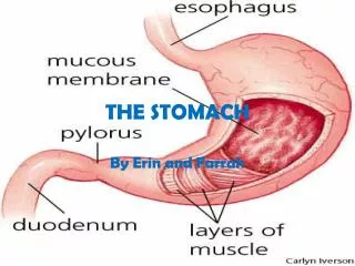
Pathology of the Stomach Michael Cohen, MD Brazos Valley Pathology
Reading Robbins and Cotran 8th ed. pp. 774-790. This presentation
Learning objectives • Etiology, pathogenesis, morphology and clinical manifestations of different types of acute gastritis, including non-erosive, erosive and hemoarrhagic. • Stress ulcers: in what context they develop. • Etiology, pathogenesis and manifestations of three types of chronic gastritis: H. Pylori, autoimmune and others (radiation, bile reflux, indwelling tubes, systemic diseases). • Brief overview of H. pylori and epidemiology of H. Pylori infection. • H. pylori associated diseases: chronic gastritis, peptic ulcer, gastric adenocarcinoma, gastric lymphoma. • Diagnosis of H. pylori infection: Urea test, serology, biopsy. • Peptic ulcer disease: Definition, epidemiology, gross and microscopic morphology, clinical manifestations and complications, etiopathogenesis. • Ménétrier disease. • Zollinger-Ellison syndrome (gastrinoma).
Learning objectives • Gastric adenocarcinoma: Morphological types (intestinal versus diffuse), epidemiology, molecular pathogenesis, staging and treatment. • Gastric lymphoma, with emphasis on MALToma. • Gastrointestinal stromal tumor (GIST): Morphology, pathogenesis, clinical features, treatment with tyrosine kinase inhibitors
Protective:Mucus, bicarbonate, regeneration, blood flow, prostaglandins Damaging:H. pylori, medications, alcohol, tobacco, ischemia, delayed emptying Mucosal defense mechanisms
Acute gastritis: Causes • Most frequent causes:NSAIDS, Alcohol, Smoking H. Pylori • Others: • CMV • Ingestion of corrosive substances • Uremia • Radiation • Chemotherapy • Gastric tubes • Bile reflux • Major stress: trauma, shock, burns
Acute gastritis: Clinical manifestations • Epigastric discomfort, gnawing/burning pain • Loss of appetite • Nausea, vomiting, bloating • Hematemesis or melena if significant bleeding present • H. pylori: Often asymptomatic
Acute gastritis: Non-erosive, Erosive, Hemorrhagic Mild acute gastritis Mild erythema and scattered tiny erosions Intraepithelial neutrophils Moderate acute erosive gastritis: Multiple erosions (red spots) Severe acute hemorrhagic gastritis
Stress ulcers (Stress-related mucosal disease) • Occur in patients suffering from shock, burn, head trauma • May lead to bleeding or perforation • Curling ulcers develop in patients suffering severe burns: Due to loss of plasma • Cushing ulcers develop in patients with closed head injury and increased intracranial pressure • Possibly due to activation of vagal nuclei • Treatment of critically ill patients with PPI or H2 blockers From Cushing’s original article describing 3 patients who died of perforated ulcer after neurosurgery. Focal hemorrhages and 3 large perforations. Pictures from: “Cushing ulcer: The Eponym and His Own.” Neurosurgery 68:1695–1698, 2011
Chronic gastritis • Cause #1:Helicobacter pylori • Cause #2:Autoimmune gastritis • Other causes: Radiation, chronic bile reflux, indwelling tubes, systemic diseases
Gram-negative, spiral shaped, flagellate, microaerophilic bacterium Lives in the gastric antrum, within the mucus layer covering epithelium Transmission: oral-oral, fecal-oral Survives in the acidic gastric environment thanks to flagella, urease, adhesins Makes proteases and phospholipases that break down mucus Immune cells can not reach bacterium Helicobacter pylori Barry J. Marshall From “The Helicobacter Foundation”
H. pylori: Epidemiology • Prevalence of H. pylori infection correlates with poverty, household crowding, lack of education, i.e. low socio-economic status • Prevalence increases with age: 50% over age 60, 20% below age 40 • In poor countries, infection is acquired in childhood
H. pylori: Associated diseases • Most infected individuals are asymptomatic (80%) • Diseases associated with H. pylori infection: • Chronic gastritis • Peptic ulcer • Gastric adenocarcinoma • Gastric lymphoma • Infected patients acquire duodenal ulcer at a rate of 1% per annum • Nearly all patients with duodenal ulcer have H. pylori Green square: uninfected people Circle: total number of infected people
H. pylori: Chronic gastritis • Dyspeptic symptoms • Endoscopy: Erythema, nodular appearance of mucosa or thickening of rugae • Pathology: • Intraepithelial neutrophils, sometimes forming pit microabscess • Lymphocytes in lamina propria with numerous plasma cells • H. PYLORI ON THE SURFACE, demonstrated by special stains
H. pylori: Chronic gastritis Antral-predominant type • H. pylori preferentially infects the antrum • Leads to hyperchlorhydria: • Increased production of gastrin from G-cells • Increased production of histamine from ECL cells • Inhibition of somatostatin release • Result:Duodenal ulcer Antral-predominant Hyperchlorhydria leads to duodenal ulcer
H. pylori: Chronic gastritisCorpus-predominant type • Atrophic Pangastritis: Infection spreads to body and fundus, causing mucosal atrophy • Progression depends on host’s and bacterial factors • Atrophic pangastritis results in hypochlorhydria • Complications: • Intestinal metaplasia/dysplasia leading to adenocarcinoma • Mucosal atrophy leading to gastric ulcer Corpus-predominant: Atrophy, hypochlorhydria, gastric ulcer, intestinal metaplasia and adenocarcinoma
H. pylori: Diagnosis • Urea test • Circulating antibodies • Esophagogastroduodenoscopy and biopsy
H. pylori: Histological identification IMMUNOHISTOCHEMISTRY SILVER GIEMSA MCMULLEN From: J Clin Pathol, 2000, 53: 756
Autoimmune gastritis – 1 • Second most frequent form of chronic gastritis • Autoimmune process targeting parietal cells and Intrinsic Factor: • Autoantibodies to parietal cells and IF present early • Injury mediated by CD4+lymphocytes, not autoantibodies • Chronic inflammation of mucosa (picture) with mucosal atrophy in body and fundus • Antrum and cardia unaffected, in contrast to H. pylori gastritis
Autoimmune gastritis – 2 • Pathophysiology: Hypochlorydria • Compensatory hyperplasia of G-cells in the antrum • Carcinoids often develop • Vitamin B12 deficiency: • Pernicious anemia(megaloblastic) • Neurologic disease • Atrophic glossitis • Intestinal metaplasia in atrophic mucosa, leading to risk of adenocarcinoma • Disease develops over decades • Diagnosis made around 60 Intestinal metaplasia: Note goblet cells
Peptic Ulcer Disease (PUD) • Lifetime risk of developing PUD is 10% for men, 4% for women • Estimated number of people with active PUD: 4 million • Duodenal:Gastric PUD is 4:1 • H. pylori associated with 65% gastric and near 100% duodenal disease From The Helicobacter Foundation
Peptic ulcer • Gross features: • Solitary, well-circumscribed mucosal defect • Flat margins and smooth base • Depth up to muscularis propria • Histology: 4 layers • 1) Superficial fibrinous exudate, 2) acute and chronic inflammation, 3) granulation tissue, 4) fibrosis • Healing leads to scar
Peptic ulcer versus ulcerated cancer • Malignant transformation of peptic ulcers is rare • Gastric cancer often presents as an ulcerated mass • “Malignant ulcers” have heaped-up margins and irregular base • Gastric ulcers should always be biopsied to rule out cancer Gastric peptic ulcer (Medscape) Ulcerated gastric carcinoma (http://web2.airmail.net/uthman/index.html)
Peptic ulcer: Location • Gastric ulcers along lesser curvature at junction between body and antrum • Duodenal ulcers located in the duodenal bulb within a few cm from pylorus on anterior surface
Peptic ulcer: Causes • In total (duodenal + gastric ulcers) 70% of patients with PUD have H. pylori • Fewer than 20% of people with H. pylori develop PUD Other etiologic factors: • NSAIDS and corticosteroids: Inhibit prostaglandin synthesis • Smoking: Reduces mucosal blood flow • Psychological stress: Increases acid production • Genetic predisposition • Cirrhosis, COPD, chronic renal failure, hyperparathyroidism: Unclear mechanisms
Peptic ulcer: Clinical manifestations • Epigastric gnawing, burning pain related to mealtimes • Onset of pain in relation to meals depends on location, duodenal versus gastric: • DU: 30 min to 3 hour after meal, relieved by antacids or food • GU: immediately before or during meal, precipitated by food • Dyspeptic symptoms: Bloating, nausea, vomiting • Weight:Loss in gastric ulcers. Gain in duodenal ulcers • Hematemesis, melena: Anemia • Mortality: Low, but 5,000 die each year of PUD’s complications in the US
Peptic ulcer: Complications • Perforation: Acute, life-threatening • Bleeding: • From small vessels: Chronic bleeding, iron-deficiency anemia • From large vessel: Hematemesis, melena, shock • Gastric outlet obstruction: Ulcers in distal stomach and duodenal bulb may cause reversible obstruction by edema or fixed stenosis by scarring • Penetration into adjacent organs, liver and pancreas From Robbins. Acute gastric perforation Free air under the diagram due to perforated gastric ulcer
Ménétrier disease (Hyperplastic Hypersecretory Gastropathy) • Hyperplasia of foveolar epithelium in body/fundus • Thickening of rugae, with cerebriform appearance • Excessive protein secretion leading to protein-losing enteropathy • Weight loss, diarrhea, edema • Due to hyperproduction of TGF-a • Increased risk of adenocarcinoma • Supportive treatment
Zollinger-Ellison syndrome • Caused by a gastrinoma, gastrin-secreting neuroendocrine tumor • Usually in the pancreas or small intestine • Leads to hyperplasia of parietal cellsand hyperchlorydria • Multiple gastric and duodenal ulcers, chronic diarrhea • Two thirds of gastrinomas are malignant but slow-growing • 25% associated with MEN I Gastrinoma: Uniform epithelial cells with round nuclei and finely dispersed chromatin (“salt-and-pepper”)
Neoplastic Pathology of Stomach • Polyps • Adenocarcinoma • Lymphoma • Gastrointestinal stromal tumor (GIST)
Gastric Polyps: 3 types A • Hyperplastic/inflammatory: Most common, chronic gastritis, unlikely malignant transformation • Fundic type: Found in FAP and PPI treatment; no malignant potential • Adenomatous: Neoplastic type, with dysplasia (low, high), malignant potential related to degree of dysplasia and size B C
Gastric Adenocarcinoma • 90% of gastric malignant tumors • Initial symptoms:Non-specific dyspepsia • Advanced stage:Weight loss, anemia • Incidence varies widely throughout the world • Screening is cost-effective in high-incidence countries • Incidence in Western countries dropped in 20th century • Migrants acquire the risk of country to where they move International Agency for Research on Cancer . Data on the annual incidence of gastric cancer in selected countries according to gender. From Fuchs, CS, Mayer, RJ, N Engl J Med 1995; 333:32.
Adenocarcinoma: 2 types Ulcerated mass Leather bottle Gland formation Signer-ring cells Intestinal type Diffuse type
Adenocarcinoma: Two types Intestinal type (90%) Diffuse type (10%) Diffusely infiltrative, with desmoplastic reaction: Linitis plastica Non-cohesive signet-ring cells Not associated with H. pylori or other risk factors Uniform incidence across countries • Bulky, ulcerated tumors • Glandular structures • Associated with known risk factors: H. pylori, atrophic gastritis, intestinal metaplasia/dysplasia, adenomatous polyps • Predominates in high-risk areas
Adenocarcinoma: Epidemiology • Epidemiology supports strong role of environmental factors • H. pylori found in 2/3 of cases • Other risk factors: Smoking and alcohol, atrophic (autoimmune) gastritis, low socioeconomic status • Dietary carcinogens: N-nitroso compounds, benzopyrene • Protective factors: Vegetables, citrus fruits containing Vitamin C and E, beta-carotene
Adenocarcinoma: Molecular pathogenesis • Intestinal and diffuse types follow different pathogenetic pathways • Diffuse type: CDH1 mutations with loss of E-cadherin function (cell adhesion) key event in diffuse type • Intestinal type: Increased risk in FAP, mutations of b-catenin(intracellular signal transducer involved in growth regulation), p53 mutations, polymorphisms of pro-inflammatory genes
Gastric adenocarcinoma: Staging and Treatment • Stage most important prognostic factor: • Depth of invasion and nodal/distant mets • Early stages, limited to mucosa and submucosa : 90% 5-year survival • Early gastric cancer can be treated with endoscopic mucosal resection • Advanced stages: 20% 5-year survival • Overall 5-year survival rate: <30% • Treatment: Surgery plus combination of radiation and chemotherapy • Trastuzumab (Herceptin), a monoclonal antibody that inactivates HER2/neu receptors, approved in 2010 for treatment of gastric adenocarcinoma
Gastric Lymphoma • GI tract is the most frequent site of extranodal lymphomas • Lymphoma represents 5% of gastric malignant tumors • Majority of lymphomas are derived from Mucosa-Associated Lymphatic Tissue(MALT) and are thus called MALTomas • It’s an extranodal marginal-cell lymphoma
MALTomas: Pathogenesis • Arise at sites of chronic inflammation, most commonly due to H. pylori • EARLY STAGE:Reversible • Antigen-dependent activation of NF-kB, a growth-promoting transcription factor, through increased expression of MLT and BCL-10 genes • Antibiotic treatment of H. pylori infection results in remission of MALToma • LATE STAGE:Irreversible • Specific translocations lead to constitutiveactivation of NF-kB • MALTomas will not regress with antibiotic treatment
MALTomas: Morphology • MALTomas are B-cell lymphomas • Dense lymphocytic infiltrate expanding the lamina propria • Neoplastic lymphocytes surround and infiltrate glands producing typical lymphoepithelial lesions • Slow-growing, eventually transform into high-grade lymphomas (diffuse large B-cell lymphoma)
Gastro-Intestinal Stromal Tumor (GIST) • GIST is the most common non-epithelial tumor of the stomach • Other non-epithelial tumors: Leiomyoma,schwannoma • All 3 look similar histologically: Spindle-cell tumors • GIST occurs in the 50s-60s • Children with GIST usually have Carney triad, i.e. gastric GIST, paraganglioma, pulmonary chondroma (non-hereditary) • Don’t make confusion with Carney Syndrome,autosomal dominant, cardiac myxoma, skin hyperpigmentation, endocrine overactivity
GIST: Pathogenesis • GIST originates from interstitial cells of Cajal, pacemaker cells in the muscularis propria • Two genes are mutated in GIST • c-KIT (AKA CD117), a tyrosine kinase receptor for stem cell factor, in 90% of cases OR • PDGFRA: Platelet-derived growth factor receptor a, a tyrosine kinase related to c-KIT, in 8% of cases
GIST: Morphology • Solitary, well-demarcated tumor covered by intact or ulcerated mucosa • Can grow to large size • Histologically, most tumors are composed of spindle-shaped cells arranged in long fascicles
DD: Gist, leiomyoma, schwannoma Leiomyoma: Positive for smooth muscle actin GIST: positive for CD-117 Schwannoma: Positive for S-100
GIST: Clinical • Anemia is the most frequent presenting sign, due to mucosal ulceration and bleeding • Vague abdominal pain, feeling of fullness, vomiting • Late stage: Metastases to the liver and peritoneal cavity • Prognosis: Size, mitoses, location • Treatment: Surgical removal, commonly by laparoscopic surgery • For unresectable and metastatic tumors, treatment with selective tyrosine kinase inhibitors: • Imatinib (Gleevec), sunitinib (Sutent)