Download
1 / 52
530 likes | 556 Views
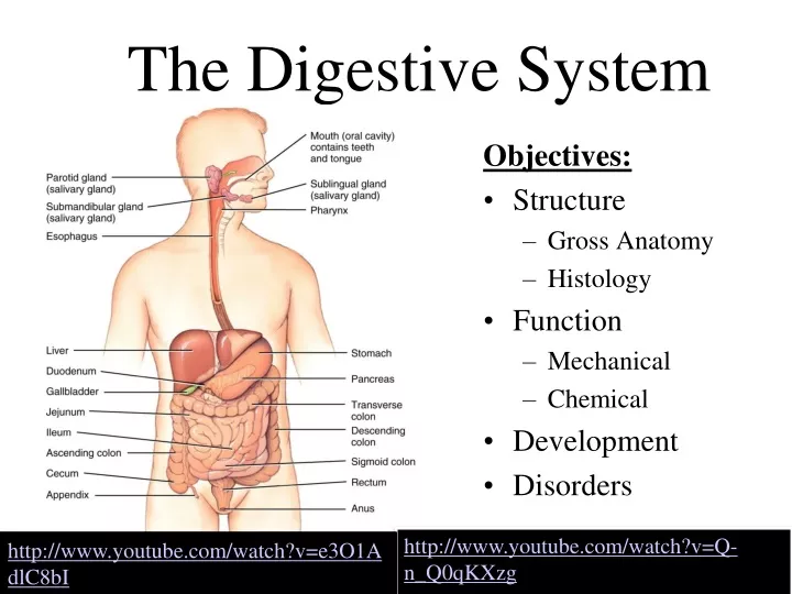
The Digestive System. Objectives: Structure Gross Anatomy Histology Function Mechanical Chemical Development Disorders. http://www.youtube.com/watch?v=Q-n_Q0qKXzg. http://www.youtube.com/watch?v=e3O1AdlC8bI. Overview of GI tract Functions. 1. Mouth - bite, chew, swallow (Mastication)

E N D
The Digestive System Objectives: • Structure • Gross Anatomy • Histology • Function • Mechanical • Chemical • Development • Disorders http://www.youtube.com/watch?v=Q-n_Q0qKXzg http://www.youtube.com/watch?v=e3O1AdlC8bI
Overview of GI tract Functions 1. Mouth- bite, chew, swallow (Mastication) 2. Saliva - part of the initial process of food digestion, enzymes in the saliva begin break down of starch and fat 3. Pharynx and esophagus- transport to… 4. Stomach- mechanical disruption; absorption of some water & alcohol 5. Small intestine- chemical & mechanical digestion & absorption 6. Large intestine- absorb electrolytes & vitamins (B and K) 7. Rectum and anus- defecation
Greater Omentum, Mesentery PURPOSE: Support/organization of bowels, Fat deposition, Immune contribution, Infection/wound isolation
Peritoneum 2 Layers: 1. Parietal peritoneum The outer layer, called, is attached to the abdominal wall 2. Visceral peritoneum The inner layer, is wrapped around the internal organs The term mesentery is often used to refer to a double layer of visceral peritoneum. There are often blood vessels, nerves, and other structures between these layers. The space between these two layers is technically outside of the peritoneal sac, and thus not in the peritoneal cavity. For our purposes the terms Mesentery and Peritoneum ought to be viewed as basically the same thing
Peritonitis • Acute inflammation of the peritoneum (the largest serous membrane in the body) • Cause • contamination by infectious microbes during surgery or from rupture of abdominal organs
Mouth • Mouths role in digestion : • Mastication of food – mechanical break down thru chewing. Mixes food with saliva • Saliva – initial chemical break down of food by enzymes, lubricates food.
Pharynx http://www.youtube.com/watch?v=Fn4pB_TJhEk&feature=related
Pharynx • Funnel-shaped tube of skeletal muscle lined by mucous membrane • Deglutition (or swallowing) is facilitated by saliva and mucus • starts when bolus is pushed into the top of the pharynx • sensory nerves send signals to deglutition center in brainstem • úvula and soft palate are lifted to close nasopharynx • larynx is lifted as epiglottis is bent to cover glottis
Cleft Palate Cleft lip and palate are birth defects that affect the upper lip and the roof of the mouth. Causes: Genetics, some viruses and toxins http://www.youtube.com/watch?v=Ehork83SreY
Esophagus • Collapsed muscular tube • In front of vertebrae • Posterior to trachea • Posterior to the heart • Pierces the diaphragm at hiatus • hiatal hernia or diaphragmatic hernia is when a piece of the GI tract pushes through the hiatus
Hernia http://www.youtube.com/watch?v=d72ezsZ46gI
Acid reflux or heart burn occurs when acidic contents of the stomach flow back up into the esophagus
Physiology of the Esophagus - Swallowing • Voluntary phase---tongue pushes food to back of oral cavity • Involuntary phase----pharyngeal stage • breathing stops & airways are closed • soft palate & uvula are lifted to close off nasopharynx • vocal cords close • epiglottis is bent over airway as larynx is lifted
Swallowing • Upper sphincter relaxes when larynx is lifted • Peristalsis pushes food down • Travel time is 4-8 seconds for solids and 1 sec for liquids • Lower sphincter relaxes as food approaches http://www.youtube.com/watch?v=jr2CuFRCsP8
Heimlich Maneuver For blocked airway http://www.youtube.com/watch?v=WleKTUaBCIw
Anatomy of Stomach • Which side is it on? • (Left) • Size when empty? • large sausage. Stretches due to rugae (a series of ridges produced by folding of the wall of an organ.) • Parts of stomach • Cardia, fundus, pylorus, pyloric sphincter • The stomach releases proteases (protein-digesting enzymes such as pepsin) and hydrochloric acid, which kills or inhibits bacteria and provides the acidic pH for the proteases to work. Food is churned by the stomach through muscular contractions of the wall and converted into chyme • Empties as small squirts of chyme leave the stomach through the pyloric valve
Muscular Structure of Stomach • Three layers of smooth muscle--outer longitudinal, circular & inner oblique • Permits greater churning & mixing of food with gastric juice http://www.youtube.com/watch?v=l4vREUUv9Lw http://www.youtube.com/watch?v=ig5KI4tswNg
SIDE EFFECTS OF GASTRIC BY PASS: • Chronic Dehydration • Difficulty swallowing • Gallstones • Hair Loss • Indigestion • Intolerance to certain foods, beverages and drugsVomiting • Change in bowel habits • (Diarrhea or loose stools and Constipation) • Kidney stones Dumping Syndrome (Combinations of weakness, dizziness, flushing and warmth, nausea and palpitation immediately or shortly after eating )
Pyloric Stenosis Belching and projectile vomiting, weight loss, dehydration
What is a peptic ulcer? A peptic ulcer is a hole in the gut lining of the stomach, duodenum, or esophagus. An ulcer occurs when the lining of these organs is corroded by the acidic digestive juices which are secreted by the stomach cells. What are the causes of peptic ulcers? While acid is still considered significant in ulcer formation, the leading cause of ulcer disease is currently believed to be infection of the stomach by a bacteria Contrary to popular belief, alcohol, coffee, colas, spicy foods have no proven role in ulcer formation. Similarly, there is no conclusive evidence to suggest that life stresses or personality types contribute to ulcer disease. http://www.youtube.com/watch?feature=endscreen&NR=1&v=K-Ao6kyoaNk
Anatomy of the Pancreas • 5" long by 1" thick • Main duct joins common bile duct from liver • Opens 4" below pyloric sphincter • Both an endocrine gland (producing several important hormones, including insulin) as well as an exocrine gland, secreting pancreatic juice containing digestive enzymes that pass to the small intestine. These enzymes help in the further breakdown of the carbohydrates, protein, and fat in the chyme.
Pancreatic Cancer The exact cause is unknown, but pancreatic cancer is more common in smokers and people who are obese. May often grow without any symptoms at first. This may mean pancreatic cancer is more advanced when it is first found. Early symptoms of pancreatic cancer include: 1. Pain or discomfort in the upper part of the belly or abdomen 2. Loss of appetite and weight loss 3. Jaundice (a yellow color in the skin, mucus membranes, or the eyes) 4. Dark urine and clay-colored stools 5. Fatigue and weakness 6. Nausea and vomiting
Liver 1. production of bile, which helps break down fats in the small intestine – sent directly to small intestine and also Gallbladder 2. production of certain proteins for blood plasma 3. production of cholesterol to help carry fats through the body 4. conversion of excess glucose into glycogen (fat) for storage 5. Storage of iron for hemoglobin (main protein of blood) 6. conversion of poisonous ammonia to urea (Urea is one of the end products of protein metabolism that is excreted in the urine.) 7. clearing the blood of drugs and other poisonous substances 8. regulating blood clotting 9. removing bacteria from the bloodstream http://www.youtube.com/watch?v=ZD_Tlu1JUoI&feature=related
Cirrhosis of the liver What is cirrhosis? Cirrhosis is a condition in which the liver slowly deteriorates and malfunctions due to chronic injury. Scar tissue replaces healthy liver tissue, partially blocking the flow of blood through the liver. Scarring also impairs the liver’s ability to control infections remove bacteria and toxins from the blood, process nutrients, hormones, and drugs, make proteins that regulate blood clotting, produce bile to help absorb fats A healthy liver is able to regenerate most of its own cells when they become damaged. With end-stage cirrhosis, the liver can no longer effectively replace damaged cells. A healthy liver is necessary for survival.
The Gallbladder The adult human gallbladder stores about 50 milliliters of bile, which is released into the duodenum when food containing fat enters the digestive tract. After being stored in the gallbladder, the bile becomes more concentrated than when it left the liver, increasing its potency and intensifying its effect on fats. http://www.youtube.com/watch?v=ECmwbwIrBI8
Gallstones are lumps or stones made up of various crystals that develop in the gallbladder or bile duct. When the chemicals in the gallbladder, cholesterol, calcium bilirubinate, and calcium carbonate are out of balance gallstones may form. What causes gallstones? Overweight/obese people, especially women, who have bulging People who have recently lost lots of weight. People whose intake of dietary fat is high. People over 60 years of age. http://www.youtube.com/watch?v=_gBEhXlg4RQ
Bile Production • One quart of bile/day is secreted by the liver • yellow-green in color & pH 7.6 to 8.6 • Components • water & cholesterol *Function: aids the process of digestion of lipids in the small intestine by emulsifying fats in partly digested food. Also “turns off” HCl and Pepsin so other digestive enzymes and bacteria can do their job
Anatomy of the Small Intestine • 20 feet long----1 inch in diameter • Large surface area for majority of absorption • 3 parts • duodenum---10 inches • jejunum---8 feet • ileum---12 feet • ends at ileocecal valve • mesentery and omentum holds the small intestine loosely in place and binds it to the posterior abdominal wall
Mechanical Digestion in the Small Intestine • Weak peristalsis in comparison to the stomach---chyme remains for 3 to 5 hours • Segmentation---local mixing of chyme with intestinal juices---sloshing back & forth
Chemical Digestion in the Small Intestine *Bile from the Liver and/or Gall Bladder flows through the common Bile Duct joining Pancreatic juice in the Hepatopancreatic duct dumping into the duodenum where it help further break down chime so that it can be acted on by intestinal bacteria and absorbed into the blood for transport to the bodies tissues.
Absorption of Water • 9 liters of fluid dumped into GI tract each day • Small intestine reabsorbs 8 liters • Large intestine reabsorbs 90% of that last liter • Absorption is by osmosis through cell walls into vascular capillaries inside villi
Anatomy of Large Intestine • 5 feet long by 2½ inches in diameter • Cecum & appendix at the beginning • Rectum = last 8 inches of GI tract anterior to the sacrum & coccyx • Anal canal = last 1 inch of GI tract • internal sphincter----smooth muscle & involuntary • external sphincter----skeletal muscle & voluntary control
Histology of Large Intestine • Muscular layer • internal circular layer is normal • outer longitudinal muscle • Appendix • A finger like vestigial structure. • Servers a minor role in bacteria growth and immunity (?) • Bacteria (e.coli) of the large intestine are responsible for the final stage of digestion. • Large intestine----absorbs electrolytes & vitamins (B and K)Electrolytes are important because they are what your cells (especially nerve, heart, muscle) use to maintain voltages across their cell membranes and to carry electrical impulses (nerve impulses, muscle contractions) across themselves and to other cells. *Digestive system normally produces about seven liters of gas per day creating 14 occurrences of flatulence per day
Blocked, Impacted, or Twisted Intestine Colon Resection: http://www.youtube.com/watch?v=07xcVlEoidU
Appendicitis Attack Symptoms Pain that intensifies when moving, taking deep breaths, coughing, or sneezing, Loss of appetite , Nausea, Vomiting, Inability to pass gas, Low fever that begins after other symptoms , Abdominal swelling, Feeling like a bowel movement will relieve the discomfort. http://www.youtube.com/watch?v=ICI6mT2U6zU Appendicitis is one of the most common causes of emergency abdominal surgery in the United States. It usually occurs when the appendix becomes blocked by feces, a foreign object, or rarely, a tumor.
Defecation Is the final act of digestion by which organisms eliminate solid, semisolid or liquid waste material (feces) from the digestive tract via the anus. Waves of muscular contraction known as peristalsis in the walls of the colon move fecal matter through the digestive tract towards the rectum. http://www.youtube.com/watch?v=XxvRbxhqoZk
What are hemorrhoids? Hemorrhoids are swollen veins in the anal canal. This common problem can be painful, but it's usually not serious. Change is diet helps, but in extreme cases surgery can be done.