Download
1 / 1
10 likes | 166 Views
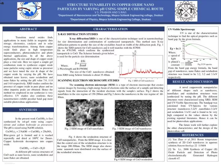
STRUCTURE TUNABILITY IN COPPER OXIDE NANO PARTICLES BY VARYING pH USING SIMPLE CHEMICAL ROUTE. UV-Visible Spectroscopy UV is one of the characterization technique to find the optical properties such as band gap, by the given formula.

E N D
STRUCTURE TUNABILITY IN COPPER OXIDE NANO PARTICLES BY VARYING pH USING SIMPLE CHEMICAL ROUTE UV-Visible Spectroscopy UV is one of the characterization technique to find the optical properties such as band gap, by the given formula. From the band gap energy formula, the band gap of nanoleaves, nano flakes and octahedron structure was found to be 3.2, 3.2 and 3.1eV respectively. Transition metal oxides finds applications in many fields in magnetic data storage, electronics, catalysis and in solar energy transformations. Among them copper oxide finds place in high temperature superconductors, photoconductive and photo thermal applications. In all the above applications, the size and shape of copper oxide plays a vital role. Here we report a simple gel combustion route to synthesize copper oxide nano particles using cupric nitrate as precursor and we have tuned the size & shape of the copper oxide by varying the pH. We have obtained nano leaves, nano octahedron and nano flakes by setting the pH value 7.0, 11.0 and 14.0 respectively. XRD studies confirm the presence of copper oxide in pure phase and no other impurity peaks are obtained. Hence the method we employed in our synthesize is very effective. UV studies also confirms these nanostructures exhibit peculiar band gap more suitable photovoltaic applications. ABSTRACT X-RAY DIFFRACTION STUDIES X-ray diffraction(XRD) is one of the characterization technique used in nanotechnology for size determination and conformation of synthesized nanoparticle. This method uses X-ray diffraction patterns to predict the size of the crystallites based on width of the diffraction peak. Fig. 1 shows the XRD pattern for CuO nanoleaves and it well matches with the JCPDS CHARACTERIZATION SCANNING ELECTRON MICROSCOPE STUDIES The scanning electron microscope (SEM) is a type of electron microscope that creates various images by focusing a high energy beam of electrons onto the surface of a sample and detecting signals from the interaction of the incident electrons with the sample's surface. Fig.2 shows the nanoflakes in the size regime of 150-200nm and Fig.3 shows the nanoleaves in the size regime of 60-80nm. SYNTHESIS RESULT AND DISCUSSION A novel copperoxide nanoparticles of different shapes such as nanoleaves, nanoflakes and octahedron structures were synthesized by tuning the pH. The different structures were characterized using XRD, SEM and UV-Visible Spectroscopy. The bandgap was calculated from UV-Spectra for various structure (nanoleaves-3.2eV ,nanoflakes-3.2eV and octahedron 3.1eV) and is found to be very high compared to the values shown by the existing reported literatures. Hence it can be used for photovoltaic applications. The future work will further explore the other characteristics and the design of the photovoltaic cell. REFERENCES In the present work Cu(OH)2 is first synthesized by sol-gel route using copper nitrate and sodium hydroxide as precursors. The pH is raised using NaOH solution. Cu(NO3)2 + 2 NaOH → Cu(OH)2 + 2NaNO3 Blue-green gel is formed and it is washed repeatedly and dried at 1000C for 2hours. Copper hydroxide decomposes into copper oxide. Cu(OH)2 → CuO + H2O At different pH various structures of CuO such as nano leaves, nano octahedron and nano flakes are obtained. 1Department of Nanoscience and Technology, Mepco Schlenk Engineering college, Sivakasi 2Department of Physics, Mepco Schlenk Engineering College, Sivakasi. V S .PRABHIN AND S.RAVI [1] Yan Zhao et al.,2011 Room temperature synthesis of 2D CuO nanoleaves in aqueous solution.Nanotechnology22 115604 [2] Yu Li., 2008 Synthesis of Copper (II) Oxide Particle and Detection of Photo electrochemically Generated Hydrogen chem46-47. file no #781588, which confirms the synthesized nanoparticle is CuO. The Scherrer formula given below is used for the particle size determination Where, λ-Wavelength and B-FWHM. The size of the CuO nanoleaves obtained from XRD using Schrrer formula is about 35-40nm. Where, c- speed of light h- planck’s constant Fig.5 UV-spectra Fig. 1 XRD of CuO nanoleaves Fig. 2 SEM image of CuO nanoflakes Fig. 3 SEM image of CuO nanoleaves Fig. 4 shows the octahedron structure of CuO nanoparticles. From the SEM image it is clear that the central axis of the octahedron structure is in the range 200-300nm. The SEM image also shows some nanorods were distributed over the surface of the octahedral structure. Fig 4 SEM image of CuO octahedron