Download
1 / 29
340 likes | 591 Views
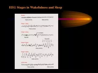
EEG Patterns During Sleep. The Neural Origin of Sleep Spindles?.

E N D
The Neural Origin of Sleep Spindles? Thalamocortical oscillations in vivo. Sleep spindle oscillations are generated by synapses in the thalamus. (A, left) Potentials recorded through a microelectrode inserted in the deafferented reticular thalamic nucleus of a cat. The arrow points to one spindle sequence. (A, right) Spindle oscillations recorded from the thalamus of a cat with an upper brainstem transection that created an isolated forebrain preparation. The figure shows two spindle sequences (the second marked by an arrow) and, between them, lower frequency (delta) waves. (B) Neuronal connections involved in the generation of spindle oscillations. (C) Intracellular recordings of one spindle sequence (see A) in three types of neurons (cortical, reticular thalamic, and thalamocortical). Ca2+, calcium ions; IPSP, inhibitory postsynaptic potential
Acetylcholine cannot, by itself, activate or shut down the neurons of the thalamus. Instead it sensitizes them. By slightly depolarizing the thalamic neurons (it does this by closing a hyperpolarizing potassium channel), the ascending system can make the thalamus more sensitive to sensory input. This corresponds to an awake, alert state
Now, although both areas are receiving acetylcholine, they have different receptors and respond in different ways. Sensory thalamus (VPL) is sensitized by acetylcholine (or "facilitated") as described above, but the reticular nucleus is inhibited by acetylcholine. But Wait, There’s MORE!
Physiological Mechanisms of Sleep: A Simplification I. The occurrence of REM Sleep: A. Controlled mostly by mechanisms located in the brainstem (particularly the pons). B. The components of REM are stimulated (turned on) by acetylcholinergic mechanisms. C. The components of REM are inhibited (turned off) by: a. Serotonergic neurons of the dorsal raphe nuclei. b. NE neurons of the locus coeruleus. c. Both NE and 5-HT serve inhibitory functions in that they prevent REM sleep from occurring when they are active.
II. The components of REM: A Desynchronized cortical EEG activity a. Possibly produced by the pons by way of the projections from the pontine nuclei (LDT=lateral dorsal tegmental nucleus & PPT= pedunculopontine tegmental nucleus) to thalamic nuclei that project broadly to various cortical areas. B. Muscular paralysis a. Possibly produced by ACh neurons (located in the dorsolateral pons near the locus coeruleus) that project caudally to magnocellular nucleus of the medial medulla and eventually to the spinal cord. C. Rapid Eye Movements a. Possibly produced by pontine nuclei (LDT and PPT) that project to superior colliculus and to the medial reticular formation.
III. Experimental evidence for mechanisms of REM: A. Lesion PPT & LDT- decrease the amount of REM sleep B. Increase number of ACh synapses in the brain - increase the amount of REM sleep C. Inhibit AChE - increase amount of REM sleep D. Inhibit AChE - shorten intervals between periods of REM sleep E. Block ACh muscarinic receptors - lengthen the interval between periods of REM sleep
IV Slow-Wave Sleep A. The nucleus of the solitary tract in the medulla is involved in slow-wave sleep. B. Slow-wave sleep may be induced by the basal forebrain region (rostral to the hypothalamus, including the pre-optic region). V. Experimental evidence for mechanisms of slow-wave sleep A. Stimulate the nucleus of the solitary tract (in the medulla) - produce EEG synchrony that looks like slow-wave sleep B. Record from the nucleus of the solitarty tract in the cat - see increased firing during slow-wave sleep. a. you do not see an increase in firing before slow-wave sleep. So, it is theorized that the nucleus of the solitary tract is involved in slow-wave sleep, but not in the induction of slow-wave sleep. C. Stimulate particular areas of the basal forebrain region - see EEG synchrony and sleep.
VI. Substances and activities that modulate sleepiness or amounts of sleep. A. Eating protein - facilitates the secretion of somatostatin - increases REM sleep. B. Eating Carbohydrates - facilitates the secretion of insulin – increases slow-wave sleep. D. Intake of or production of (due to physical activity) muramyl peptides – (note: this is interesting because serotonin can serve the same function) facilitates the release of interleukin 1 in the brain - increases slow-wave sleep. E. Intake of tryptophan rich foods - facilitates sleep induction. Caution tryptophan supplements taken in high doses will distort sleep patterns.
DYSSOMNIAS (Disorders of sleep or wakefulness) • Common Circadian Rhythm Sleep Disorders • Advanced Sleep Phase Syndrome (ASPS) - bright light therapy • Delayed Sleep Phase Syndrome (DSPS) • Time Zone Change Syndrome - Jet Lag • Intrinsic Sleep Disorders (Disorders that either originate or develop from within the body) • Hypersomnia (Excessive Sleepiness) • Insomnia • Narcolepsy • Obstructive Sleep Apnea (OSA) • Central Sleep Apnea • Periodic Limb Movement Disorder (PLMD) • Restless Legs Syndrome (RLS) • Extrinsic Sleep Disorders (Disorders that either originate or develop from causes outside the body) • Nocturnal Eating (Drinking)Syndrome (Sleep Eating)
PARASOMNIAS (Disorder of arousal, partial arousal or sleep stage transition) • Arousal Disorders (A parasomnia disorder presumed to be due to an abnormal arousal mechanism) • Confusional Arousals (Sleep inertia) • Somnambulism (Sleepwalking) in adults • Somnambulism (Sleepwalking) in children • Sleep Terrors in children • Sleep Terrors in adults • Sleep-Wake Transition Disorders • Nocturnal Leg Cramps • Rhythmic Movement Disorder • Sleeptalking
Parasomnias Usually Associated with REM Sleep • Nightmares • Sleep Paralysis • REM Sleep Behavior Disorder (RBD) • Other Parasomnias • Infant Sleep Apnea • Primary Snoring • Sudden Infant Death Syndrome (SIDS) • Sleep Bruxism (tooth grinding or tooth clenching) • Sleep Enuresis (Bedwetting) in children
Proposed Sleep Disorders • Short Sleeper • Long Sleeper • Subwakefuness Syndrome • Fragmentary Myoclonus • Sleep Hyperhidrosis (Excessive sweating) • Menstrual-Associated Sleep Disorder • Pregnancy-Associated Sleep Disorder • Terrifying Hypnogogic Hallucinations • Sleep-Related Neurogenic Tachypnea • Sleep-Related Laryngospasm • Sleep-Choking Syndrome
SLEEP APNEA Central sleep apnea. During waking, the respiratory oscillator of the medulla receives tonic drive from other neural structures and can respond to voluntary and metabolic signals to change breathing pattern. Muscle tone keeps the oropharynx open to the flow of air. In NREM sleep, central drive decreases, and the rate and depth of ventilation fall. If the airway collapses, prolonged apnea (lack of breathing) may result. During REM sleep, activation of pontine generator neurons drives the respiratory oscillator, and desynchronization may lead to breathing efforts that are too frequent or strong (hyperpnea) or that stop. During REM sleep the oscillator also becomes unresponsive to metabolic signals
What are the symptoms of Narcolepsy? -excessive sleepiness or sudden muscle weakness cataplexy (a sudden loss in muscle tone and deep tendon reflexes leading to --muscle weakness, temporary paralysis or a complete postural collapse. Cataplexy is usually brought on by an outburst of emotion - notably laughter, anger or startle.) -hypnagogic hallucinations -automatic behaviors (like driving home and not remembering how you got there!) -disrupted major sleep episode (disruption of the longest sleep episode that occurs on a daily basis).
EEG recording during sleep shows one or more of the following: -The onset of sleep is less than 10 minutes -The onset of REM sleep is less than 20 minutes and -A Multiple Sleep Latency Test (MSLT) that demonstrates an average sleep onset of less than 5 minutes Physiology shows the following: -HLA typing demonstrates DR2 positivity (Blood contains markers for narcolepsy) - Lack of hypocretin – 1 in CSF - Perhaps due to a destruction of the hypocretin (orexin) cells in the lateral hypothalamus or a point mutation in the gene coding for hypocretin.