Download
1 / 48
480 likes | 584 Views
Skeletal System. Chapter 7 Pages 130-175. Partner up!. Both you and your partner should each have 13 note cards. Pick a side (right or left)—these will be the terms that you write on your cards.

E N D
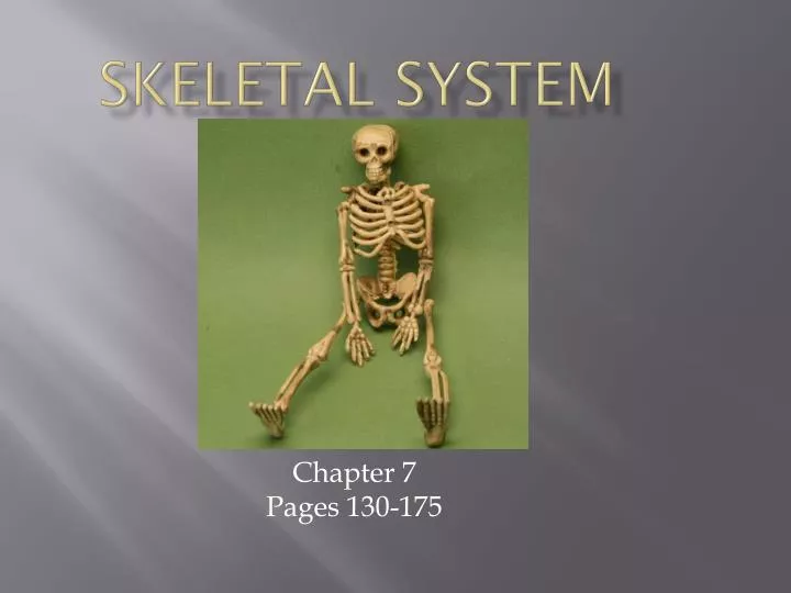
Skeletal System Chapter 7 Pages 130-175
Partner up! • Both you and your partner should each have 13 note cards. • Pick a side (right or left)—these will be the terms that you write on your cards. • When both of you have finished recording the terms, try your best to match them up (they’re not in the correct order)! • You will be quizzed on these terms in class next time!
Write these terms, one per card:Left: Right: • ax- • -blast • carp- • -clast • condyl- • corac- • cribr- • crist- • fov- • glen- • meat- • odont- • poie- • Axis • Break • Bud • Crest • Crow’s beak • Joint socket • Knob • Make/produce • Passage • Pit • Sieve • Tooth • Wrist
Let’s see how you did! • Also, don’t forget to write the correct corresponding term on the other side of your note card to study from!
Ch. 7 Wordbytes: • ax- = axis • -blast = bud • carp- = wrist • -clast = break • condyl- = knob • corac- = a crow's beak • cribr- = sieve • crist- = crest • fov- = pit • glen- = joint socket • meat- = passage • odont- = tooth • poie- = make/produce
Classification of Bones • There are 206 bones in the body grouped into two divisions: • Axial relating to the head, neck, and trunk—protect, support, or carry other body parts • E.g. head, hyoid, ribs, sternum, & vertebrae • Appendicular bones of upper & lower limbs plus girdles that connect them—help us get from place to place and to manipulate our environment
Types of Bones • Long bones- longer than wide, has shaft + 2 ends, includes all bones of limbs (- patella, wrist, ankle) • e.g. thigh, leg, arm, forearm, fingers & toes • Short bones- almost cube shaped • Most wrist & ankle bones, sesamoid bones • Flat bones- thin & extensive surface • e.g. Cranial bones, sternum, ribs & scapulae • Irregular bones- don’t fit above • e.g. vertebrae, some facial bones, hip bone
Bone Function • Support • Protection • Movement • Mineral homeostasis • Esp. Calcium and Phosphate • Blood cell production • Hematopoiesis in red bone marrow • Triglyceride Storage • In yellow bone marrow
Bone Textures • Compact bone dense outer layer—looks smooth and solid • Spongy bone internal layer—honeycomb of small, needle-like pieces = trabeculae • Open spaces filled with red/yellow marrow
Gross Structure Parts of a long bone: • Diaphysis shaft; the long, cylindrical, main portion of the bone • Medullary cavity hollow space within diaphysis that contains yellow bone marrow • Epiphysis the distal and proximal ends of the bone • Articular (hyaline) cartilage covers joint surface to cushion and absorb stress • Metaphysis • Mature bone = where diaphysis joines epiphysis • Growing bone = contains epiphyseal plate to allow diaphysis to grow
Membranes: • Periosteum dense irregular tissue that surrounds bone surface where not covered by articular cartilage • Endosteum thin membrane lining medullary cavity that contains bone-forming cells
Throw me a bone! • Come up and pick out your own to label! • Using a pen, please label the following: • Diaphysis • Metaphysis x2 • Epiphysis x2 • Type of bone • Outside texture of bone • On the back of the bone, record the 6 functions of bones!
Parts of short, irregular, and flat bones • Thin plates of periosteum-covered compact bone on the outside, endosteum-covered spongy bone within • Not cylindrical = no epiphyses • Bone marrow (between trabeculae), but no marrow cavity. • Flat bones have diploë = internal layer of spongy bone
Hematopoiesis • Newborns medullary cavity and all areas of spongy bone contain red bone marrow • Adults medullary cavity extends into epiphysis, and little red marrow present in spongy bone of long bones • Blood cells produced in head of femur and humerus (long), sternum (flat), hip (irregular) • Yellow can revert to red if needed
Microscopic Structureof Compact Bone • Osteon structural unit of compact bone=group of hollow tubes (lamellae) of bone matrix • Haversian canal runs through the center of the osteon; contains blood vessels and nerve fibers to support cells • Volkmann’s canals connect blood and nerve supplies
Microscopic Structure • Matrix= • 25% water, 25% collagen fibers (flexibility and strength), 50% crystallized mineral salts (hardness/resist compression) • Osteogenic cells in periosteum Osteoblasts secrete collagen fibers- • Build matrix and become trapped in lacunae • Become osteocytes- maintain bone • Osteoclasts (“bone breakers”) • Digest bone matrix for normal bone turnover
Compact Bone Structure • Lacunae- “lakes”; contain osteocytes • Canaliculi- little canals that allow nutrient flow from canals and between osteocytes
Spongy Bone • Units containing trabeculae to resist stress—only a few cell layers thick • Spaces between trabeculae often contain Red Marrow • No osteons but include lacunae & canaliculae
Figure 6.2b Pg. 179 Fig. 6.5
Bone Development p. 133-135 • Ossification • 1. Formation of Bony Skeleton • 2. Intramembranous Ossification • 3. Endochondral Ossification • 4. Postnatal Bone Growth • 5. Growth in length and width
Bone Development • Osteogenesis and Ossification indicate the process of bone tissue formation • In embryos this leads to formation of the bony skeleton • Bone growth goes on until early adulthood • Ossification in adults is mainly for remodeling and repair of bones
Formation of Bony Skeleton • Mesenchyme model - replaced with bone 1. Intramembranous - Bone forms directly in mesenchyme layers (membrane like) -membrane bone 2. Endochondral - forms within hyaline cartilage developed from mesenchyme- cartilage or endochondral, bone
IntramembranousOssification pg. 133 (Fig. 7.4) • Development of ossification center- • Cells differentiate=> osteogenic=> osteoblasts • Osteoblasts secrete organic matrix • Calcification- cells become osteocytes • In lacunae they extend cytoplasmic processes to each other • Deposit calcium & other mineral salts • Formation of trabeculae- spongy bone • Blood vessels grow in and marrow is formed • Mesenchyme=> periosteum • Bone Collar of compact bone forms and red marrow appears
EndochondrialOssificationpg. 134 Figure 7.5 • Uses hyaline cartilage “bones” • Cartilage must break down as process proceeds • Primary Ossification Center • Center of hyaline cartilage shaft • Blood vessels fill perichondrium • The mesenchymal cells specialize into osteoblasts
Endochondrial Ossification • Bone Collar forms around diaphysis of hyaline cartilage • Cartilage in center of diaphysis calcifies and forms cavities • Periosteal bud invades internal cavities spongy bone forms • Diaphysis elongates and a medullary cavity forms • The epiphyses ossify
Figure 6.8 Fig. 7.5 pg. 134
Postnatal Bone Growth • Length- chondrocytes in the epiphyseal plate divide and increase cartilage layer – zone 1 - Growth • On diaphyseal side they die and are replaced by bone - zone 2 – transformation • Eroded by osteoclasts, then quickly covered with bone matrix, forming spongy bone – zone 3 – osteogenic • Stops during adolescence • Periosteum supports surface growth for thickness – bones widen as they lengthen
Hormonal Regulation of Bone Growth • A symphony of hormones regulate bone growth during youth • Infancy and childhood – growth hormone – released by pituitary gland this is regulated by Thyroid hormones (T3 and T4) • Puberty – testosterone and estrogen promote adolescent growth spurt • Excess or deficits of hormones result in abnormalities – gigantism or dwarfism
Bone HomeostasisRemodeling & Repair • 5 to 7% of bone mass recycled weekly • ½ gram of calcium may enter or leave the skeleton each day • Spongy bone replaced every 3 to 4yrs. • Compact bone every 10 years
Bone Remodeling = Bone deposits and bone resorption • Bone deposit & bone resorption (removal) occur at the periosteal and endosteal surfaces. • Coordinated by “packets” of osteoblasts called remodeling units • Occurs where bone is injured or added strength is needed • Optimal bone deposit = diet of protein, adequate minerals (Ca, Mg, P), and Vitamins A, C, D
Bone resorption • Done by osteoclasts= large multinucleate cells • Osteoclasts move along bone digging pits, resorption bays, breaking down bone matrix • The ruffled border secretes lysosomal enzymes and acids • These break down the organic matrix and calcium salts into a solution that can be transported into the interstitial fluid then the blood
Classification of Fractures • 1. Nondisplaced or displaced • 2. Complete (bone in two or more pieces) or Incomplete (partial break (crack)) • 3. Orientation of break – linear or transverse • 4. Closed (simple)- not through skin or Open (compound)- broken ends break skin
Types of Fractures pg. 136-7 • Greenstick incomplete break (children—more matrix = more flexibility) • Fissured incomplete longitudinal break • Comminuted bone fragments into 3 or more pieces (elderly—brittle bones) • Transverse complete, with break at right angle to axis of bone
Types of Fractures pg. 136-7 • Oblique occurs at an angle other than a right angle • Compression bone crushed (porous bones—extreme trauma) • Spiral ragged break from twisting forces (sports fracture) • Epiphyseal separates along epiphyseal plate • Depressed broken bone pressed inward (skull fracture)
Repair of Fractures • Involves 4 major phases • 1. Hematoma formationmass of clotted blood forms at the fracture point • 2. Fibrocartilaginous callus formation for debris cleanup, bone reconstruction, bone splint • 3. Bony callus formation 3-4 weeks after injury to 2-3 months later • 4. Bone Remodelingexcess material removed, compact bone reconstructed
Homeostatic Imbalances of bone • Imbalances between bone formation and bone resorption underlie nearly all diseases that influence the adult skeleton • 1. Osteomalacia – inadequately mineralized bones • 2. Rickets – analogous disease, but in children, very severe
Homeostatic Imbalances of bone • 3. Osteoporosis – refers to a group of disorders in which bone resorption outpaces bone deposit • Occurs most often in aged, most often in postmenopausal women • Traditionally treated with calcium and vitamin D supplements
Figure 6.28 Pg. 190 – fig. 6.14 – similar photo
Homeostatic Imbalances of bone • 4. Paget’s Disease – excessive bone formation and breakdown • Often discovered by accident when X rays taken for another reason • “Spotty” weakening of bone • Can affect any part of the skeleton • Spine, pelvis, femur & skull most commonly affected
Exercise & Bone Tissue • Bone strengthened in response to use • Reabsorbed during disuse • e.g. Bone loss during bed rest, fractures in cast, astronauts with no gravity
Compact bone ground Osteon (with osteocyte inside) Pg. 179 Fig. 6.5
Bone decalcified Periosteum Spongy bone Compact bone Red marrow (spaces) Skeletal Muscle Blood vessels