Download
1 / 69
800 likes | 1.31k Views
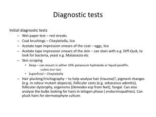
Diagnostic tests. Reasons for medical tests To confirm or exclude a proposed diagnosis To screen for disease To screen for the presence of risk factors To monitor the course of al illness To monitor the effect of treatment. Categories of patients.

E N D
Diagnostic tests • Reasons for medical tests • To confirm or exclude a proposed diagnosis • To screen for disease • To screen for the presence of risk factors • To monitor the course of al illness • To monitor the effect of treatment
Categories of patients • Those with signs and symptoms of a specific illness or condition and the test will either confirm or exclude a diagnosis. • Broad screening tests for patients who have non specific symptoms or present with vague signs of illness eg FBE. • Screening tests for patients with no signs or symptoms . The test aims to detect the presence of disease before it has manifested (eg PSA) or identify risk factors (eg elevated cholesterol).
pathology • Is the collection of specimens such as blood, tissue, body fluids and using laboratory tests to find the abnormal values/ structure etc. • Histology of tissue • System / organ functions • Immunity , infection, autoimmunity • Genetics- chromosomal DNA • Drug monitoring • Cancer markers
Diagnostic imaging • X –Ray- with or without contrast, video Scans • .
MRI- combination of large magnets, radiofrequencies and a computer
Ultrasound – high frequency sound waves and a computer are used to create an image.
Scans-Nuclear – small amounts of radioactive substances are used.
scopes • Many of the scopes today are fiber optics which allows the catheter to be flexible • These instruments can be inserted into organs and cavities. • The structure/s are either observed directly or viewed on a screen. • Dyes and X-Rays can also be used
Respiratory system diagnostic tests • http://mips.stanford.edu/research/quon/ • Bronchoscopy • A fiber optic endoscope is inserted into the bronchus • The patient is fasted and sedated
bronchoscopy • Tumors or bronchial cancer • Airway obstructions and or strictures • Inflammation and infections such as tuberculosis, pneumonia, or fungal or parasitic lung infections. • Interstitial pulmonary disease • Persistent cough or haemoptysis • Biopsy of tissue or collection of other specimens, such as sputum • Vocal cord analysis
Bronchoscopy -therapeutic • Removal of secretions, blood, mucus plugs, or polyps (growths) to clear airways. • Control bleeding in the bronchi • Removal of foreign objects or other obstructions • Laser therapy or brachytherapy (radiation treatment) for bronchial tumors. • Stent placement ( a device used to keep the airway open) • Draining of an abscess
Bronchoscopy complications • Bleeding • Infection • Bronchial perforation • Bronchospasm or laryngospasm • Pneumothorax
Lung biopsies • Types • Needle biopsy- under CT or fluoroscopy guidance • Transbronchial biopsy- via bronchoscope • Thoracoscopic biopsy or video – assisted thoracic surgery (VATS) biopsy- after a general anesthetic is given, an endoscope is inserted through the chest wall into the chest cavity. • In addition therapeutic procedures such as the removal of a nodule or other tissue lesion my be performed. • Open biopsy- after a general anesthetic is given, the physician makes an incision in the skin on the chest and surgically removes a piece of lung tissue.
Lung perfusion and / or ventilation scans • A dye is either • Injected into a vein and the blood flow to the lungs and the alveoli is observed (perfusion) • This test shows pulmonary embolism or • Inhaled into the lungs to assess the ventilation capabilities of the lungs.
Thoracentesis • Is the removal of effusion from the pleural space for • Diagnosis purposes- infection, malignancy • Therapeutic purposes – remove excess fluid, to re-expand the lung • Performed under local anesthetic • Post Procedure check • Vital signs especially respiratory rate and cough • Watch for signs of distress , shock and bleeding • dressing
bloods • FBE • U&E’s • Tropinin levels • Group and cross match
FBE • Haemoglobin (Hb) • Red Cells • Number • Shape – eg sickle cell, spherocytes, pencil cells, ovalocytes • Size – normo- micro- macro-cytic • Colour –normo- hypo- chromic • White cell count and differentiation • Platelet count
Urea and electrolytes • Urea is formed in the liver from the by products of protein metabolism. • The levels will be raised if the kidney filtration rate is less than 50 % of normal. • Other causes of raised urea are • Diet high in protein • Loss of salt and water eg vomiting , diarrhoea • Decreased blood flow to the kidneys eg CCF • Low levels can be due to • Severe liver damage • Poisoning
electrolytes • Acid –base balance • Normal 7.4 • Acidic 7.36 • Alkaline 7.44 • Water sodium balance • Electrolytes • Sodium potassium • Chloride Bicarbonate • Calcium Magnesium
Tropinin • Tropinin is a part of muscle • There are two types that are found only in cardiac muscle. • If the level of these is raised then there has been some damage to the myocardium –AMI • There may be mild elevation in severe unstable angina
Bone marrow biopsy • Reasons for doing • Diagnose certain conditions • Assess the stage or progression of certain conditions • monitor treatment of certain conditions • Procedure • Intravenous (IV) sedation • Local aesthesia • Complications • Bleeding • Infection
Cardiac catheter • A cardiac catheter is performed to view the obstructed coronary blood vessels. • The patient is awake but sedated. • A dye is injected to show the blood vessels. • Complications • Bleeding • Angina • AMI
Electrocardiograph - ECG • Views the conduction of the heart • The tracing shows PQRST formation • P wave = depolarization of the atria • QRS = depolarization of the ventricle • T wave = depolarization of the ventricle
Echocardiograph • Uses ultrasound and computer technology to crate an image of blood flow through the heart. • It can be done through the chest wall or via the oesophagus ( posterior view of the heart). • If done via the oesophagus the patient is to be fasted and sedated.
Electrocardiograph - ECG • One small square is 0.04 seconds. • One large square ( 5 small squares) is 0.2 • Damage or malfunction of the heart can be observed in an ECG. • Also the heart can be calculated.
Doppler • A Doppler uses sound waves to study the flow and rate of blood through vessels . • It can depict alterations to the flow of blood through vessels (blockages)
Angiograms and venograms • Dyes are injected into arteries or veins to highlight the flow of blood through the vessels. • The vessels can be anywhere in the body. • http://www.ohioheartandvascular.com/cvprocedures/cardiac-catheterization.php • (great site to view the dye in arteries of heart)
Lumbar puncture (spinal tap) • Reasons for performing • Meningitis and encephalitis • Metastatic tumors and central nervous system tumors. • Syphilis • Bleeding (hemorrhaging) in the brain and spinal cord. • Multiple sclerosis. • Guillain-Barre, a demyleinating disease involving peripheral sensory and motor nerves
Lumbar puncture • Post procedure • Lay flat for 4-6 hours • Neurological observations and • Check wound site (dressing) • Lumbar puncture headaches typically begin within two days after the procedure and persist form a few days to several weeks or months. • Complications • Infection • Bleeding or CSF discharge from site of entry • Numbness to legs and lower back pain
myelogram • During a lumbar puncture a dye is injected into the subarachnoid space • Reasons for procedure • Herniated discs • Spinal cord or brain tumors • Ankylosing spondylisis • Bone spurs • Arthritic discs • Cysts – benign capsules that may be filled with fluid or solid matter tearing away or injury of spinal nerve roots • Aracnoiditis – inflammation of arachnoid mater. • http://video.about.com/backandneck/Myelography.htm
Electroencephalograph (EEG) • Observes the electrical activity of the brain. • Reasons for procedure • Diagnosis of epilepsy or brain injury • To assess conditions and diseases that affect the brain.
Urinary system diagnostic procedures • Glomerular filtration rate measures the volume of blood filtered by the Glomerular membrane to form the Glomerular filtrate . • Blood flow • Blood pressure • The number of functioning glomeruli • Permeability of the glomerular membrane • Back pressure in the tubules. • Still most used to determine kidney function. • Declines as we get older
Glomerular filtration rate (GFR) • is the amount of filtrate formed by both kidneys per minute; in a normal adult, it is about 125 ml/minute. This amounts to 180 liters per day.
Serum urate • Uric acid is the breakdown of purine components (guanidine and adenine) of the nucleic acids • 1/3 derived from the diet (meat and meat products) • 2/3 derived from turnover of body cells • Can also be measured from 24 hour urinary specimen.
urea • Urea is the end product of protein metabolism. • Urea levels rise with • High protein diets • Excessive tissue breakdown • GI bleeding