Download
1 / 23
230 likes | 363 Views
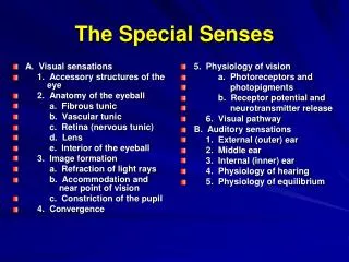
The Special Senses. External Anatomy of the Eye. Lacrimal Apparatus of the Eye. Anatomy of the Eyeball. Accessory structures of the Eye from a sagittal view. Detail view of the anterior anatomy of the eye. Photomicroscopic view of the Histology of the Eye

E N D
Photomicroscopic view of the Histology of the Eye showing the location of the central fovea
Light Refractory • Pathway: • Bulbar Conjunctiva • Cornea • Aqueous Humor • Lens • Vitreous Humor • Ganglion Cell Layer • Inner Synaptic Layer • Bipolar Layer • Outer Synaptic Layer • Photoreceptor Layer
Abnormalities of • The Eye: • Myopic - • nearsighted • Hypermetropic - • Farsighted • Presbyopia - • age-related failure of • lens to accommodate • Astigmatism - • Distorted vision due to • irregular-shaped lens or • cornea • Color Blindness - • genetic defect that • causes dysfunction of • cones
Accommodation of the Lens for near vision • Ciliary muscles contract • Ciliary body pulls forward and inward • Tension on suspensory ligaments of lens is decreased • Lens becomes thicker (rounder) due to its elasticity • Pupils constricts
Accommodation of the Lens for far vision • Ciliary muscles relaxes • Ciliary body returns to its resting state, backward and outward • Tension on suspensory ligaments of lens is increased • Lens becomes thinner (flatter) due to its elasticity • Pupils dilate
Anatomy of Rods and Cones
Visual Pathway • Cones • Bipolar neurons • Ganglion cell’s axon forms the optic nerve • Optic nerve to the Optic Chiasm • Optic tract • Lateral geniculate nuclei of the thalamus • Optic Radiations • Primary visual areas of the occipital lobes