Download
1 / 1
20 likes | 163 Views
3-D Cylinders. Control. Tumor. M. D. A NDERSON Cancer Center. Computed Tomography (micro-CT) Dianna D. Cody, Ph.D., Evan Johnson and Daniel Leventhal Small Animal Cancer Imaging Research Facility. Achondroplasia Gene Therapy Study

E N D
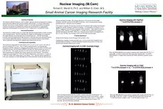
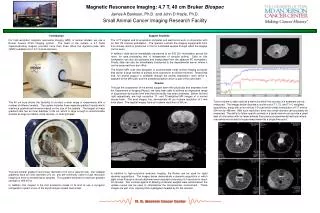
3-D Cylinders Control Tumor M. D. ANDERSON Cancer Center Computed Tomography (micro-CT) Dianna D. Cody, Ph.D., Evan Johnson and Daniel Leventhal Small Animal Cancer Imaging Research Facility Achondroplasia Gene Therapy Study The Dental Branch of the University of Texas Health Science Center conducted a study led by Dr. Pauline Duke seeking a gene therapy treatment for Achondroplasia, the most common form of dwarfism. Dr. Duke’s team modified a gene in mice, hoping that modifying this gene would cause Achondroplasia. To prove this hypothesis, a control group of natural mice and a mutated group were imaged using both conventional X-Ray and MicroCT. The MicroCT produced 3-D images of the skulls that showed something that the original investigators had not considered. The dome of the skull in the mutant mice was significantly less dense than the control group, which cannot be seen in X-Rays. MicroCT images were also used to measure the length of some bones. Ongoing Projects and Results NASA Bioreactor Skull Analysis The Dental Branch of the University of Texas Health Science Center conducted a study led by Dr. Pauline Duke where embryonic limb bud cells were grown in NASA’s Synthecon zero-G bioreactor. These cells differentiated into cartilage cells, some of which were fixed for histological study. Others were implanted in two mm circular defects in mouse skulls. The MicroCT was used to image the skulls with the implanted cells, and a control group with defects, but no implanted cells. Imaging proved that the defect had been completely filled in in the implanted mice, but not in the control mice. Furthermore, the CT scan showed that the filled-in defects had the same mineral density as the surrounding bone. Overview of Modality The EVS Corp RS-9 In Vivo MicroCT Scanner is designed to image small laboratory animals, such as mice and rats. It is an ideal instrument for biomedical research laboratories to non-destructively acquire 3-D images of both in vivo and ex vivo specimens. Instrument Description EVS Corp’s RS-9 MicroCT utilizes a cone-shaped X-Ray beam and a two dimensional detector, the latest in CT technology. Cone beam CT takes an entire image in one rotation of the gantry, without any movement necessary in the axial direction. This results in a true volume image instead of the “slices” commonly associated with CT in the past. Scans can be taken with resolution as high as 27 microns, with 90 micron resolution possible for the shortest scan. A Defect Healed Defect X-Ray Source Figure 3 (left) A control mouse skull with the defect still present. (right) A mouse skull with a healed defect due to implanted cartilage cells. Note MicroView’s line segment function is used to measure the diameter of the defects, and even though the defect is filled in on the mouse on the right, the defect diameter is slightly larger. 2-D Detector Vitamin D Bone Tumor Treatment Dr. Sara Peleg conducted a study analyzing the effectiveness of vitamin D in protecting against local bone loss in tumor-bearing bones. The right distal femur of each mouse had been inoculated with osteoclastic prostate cancer cells (PC3), which proceeded to destroy the surrounding bone tissue. The left hind leg of each mouse was not treated. A vitamin D analog treatment was applied systemically to protect against local bone loss, at two doses (4 and 10 g/kg) to 5 mice per dose group. Both legs were disarticulated and scanned in the 25 m isotropic voxel mode. Specific volumes of bone tissue were analyzed with respect to bone mineral density in the tibia of each leg. Significant differences (p=0.002) were observed between paired bone samples (tumor vs non-tumor bearing bones). Sampling errors (17-25% variation in repeated measures) could have somewhat obscured significance. The ability to re-orient the 3D data sets to a standard position prior to volume analysis is expected to improve this result. Wild type mouse 3D and sagittal view (left). Mutant mouse 3D and sagittal view (middle). Illustration of the plane location tool in MicroView (right). Figure 1 (left) A n illustration of the volume cone beam system. (right) The actual EVS RS-9 Micro-CT. Lung Tumor Imaging When attempting to study the effect of new drugs, researchers often employ animal models who already have the disease. Unfortunately, in most cases, not every animal possesses the disease to the same degree. Dr. Oh and Dr. Kurie are attempting to employ the scanner to image live animals and assess the lung tumor density in each animal before the start of treatment. These images can then be matched up with post-sacrifice histological sections to assess the effectiveness of the treatment. This application of the MicroCT is not yet perfected, but is improving with experience and upgrades, including a respirator and infusion pump for contrast dye. Technical Capabilities The RS-9 is capable of both In Vivo and Ex Vivo scanning. Ex Vivo scans often utilize the high resolution scanning mode, in which a scan takes approximately 80 minutes. However, a low resolution In Vivo scan can be completed in around 20 minutes. To perform an In Vivo scan, the veterinary team anesthetizes the animal using isoflourane. While an anesthetized animal remains relatively still during the scan, imaging of the lungs is difficult because of motion artifact. Hopefully, the use of a newly-acquired respirator synchronized with the scanner will allow imaging of the lungs exclusively in the inflated position. Cross-sectional views of both control and tumor-bearing bones (left). Examples of MicroView’s system for measuring bone density (right). A 3D cylinder is placed over the area to be measured. Average bone density within the cylinder can then be displayed. Figure 2 A rat is anesthetized for a scan. (left) View before the bed is moved into scanner. (right) View of the rat during the scan. The scan lasts approximately 20 minutes. Various views from a volume created by a 20 minute in vivo scan of a rat with no contrast dye or respiratory gating. With hardware upgrades and use of secondary software packages, the quality of these images are expected to improve dramatically.