Download
1 / 33
330 likes | 542 Views
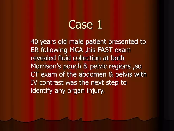
Case 1. 40 years old male patient presented to ER following MCA ,his FAST exam revealed fluid collection at both Morrison's pouch & pelvic regions ,so CT exam of the abdomen & pelvis with IV contrast was the next step to identify any organ injury.

E N D
Case 1 40years old male patient presented to ER following MCA ,his FAST exam revealed fluid collection at both Morrison's pouch & pelvic regions ,so CT exam of the abdomen & pelvis with IV contrast was the next step to identify any organ injury.
CT exam of the abd& pelvis with IV contrast revealed: • Stranding &hematoma of the mesentery • Minimal fluid collection noted at the right paracolic gutter& pelvic regions • Bowel loops near the site of mesenteric stranding are seen slightly distended.
Diagnosis Mesenteric injury
This patient was managed conservatively for one week and he had uneventful recovery.
CASE 2 23 years old male, presented to ER after his belly had been squeezed between a machine and the wall, he had positive FAST exam for free fluid collection at Morrison'sPouch, splenorenal& pelvic regions & so CT exam of the abdomen and pelvis following IV contrast was the next step.
CT exam of the abdomen & pelvis following IV contrast revealed: • extraluminal free air(multiple small air bubbles) are noted throughout the whole abdomen. • intramural air is also noted (this with extraluminal air strongly suggest full thickness rather than partial thickness injury). • free intrabdominal, pelvic& interloop fluid collection is also noted. • bowel wall thickening. • bowel wall are also seen enhanced. • Mesenteric stranding is also noted.
Diagnosis Small Bowel Trauma
At surgery the patient had atear at the proximal jejunum where the bowel loops were distended at and near the site of the tear; resection anastomosis of the affected bowel loop was done.
Case 3 • 15 years old male patient, he was hit by amotorcycle, the patient had negative FAST exam at the same day of the accident and so he was discharged; • The next day he presented again to ER with vomitting & abdominal pain .
US didn't reveal any fluid collection though a heterogenous mass appeared infront of the right kidney ,to the right of the pancreas,posterior to the gallbladder) at the typical site of the second part of the duodenum.it appears with hyperechoic periphery &anechoic center) so non enhanced CT abdomen & pelvis was performed then CT following oral & iv contrast,that confirmed the mass which appears heterogenous also at the CT with enhancing periphery with internal extensions
Diagnosis DUODENAL HEMATOMA
Blunt trauma Bowel & Mesenteric injury • Detection of bowel and mesenteric injury can be challenging in patients after blunt abdominal trauma. Early diagnosis and treatment are critical to decrease patient morbidity and mortality. • Computed tomography (CT) has become the primary modality for the imaging of these patients .
Hemoperitoneum detected with ultrasonography is no longer an indication for exploratory laparotomy in a stable patient. More emphasis is now placed on nonsurgical management of ,spleen and renal injuries. • The concurrent presence of significant bowel or mesenteric injury, however, would make conservative treatment inappropriate and necessitates exploratory laparotomy. Therefore, greater sensitivity and specificity of imaging studies are demanded for these types of injury .
CT findings of bowel trauma: • Bowel Discontinuity (Definite sign). • Extraluminal Oral Contrast Material (pathognomonic of bowel injury). • Extraluminal Air (pathognomonic of bowel injury). • Intramural Air. • Bowel-Wall Thickening. • Bowel-Wall Enhancement . • Intraperitoneal and Retroperitoneal Fluids. • Mesenteric foci of fluid, air, or fat stranding may be secondary to bowel injury alone
Findings Specific to Mesenteric Injury • Mesenteric Extravasation (pathgnomonic). • Mesenteric Vascular Beading. • Abrupt termination of Mesenteric Vessels. • Mesenteric Infiltration. • Mesenteric Hematoma
NB: If there is no other explanation for intraperitoneal fluid,bowel or mesenteric injury should be considered.
Duodenal Hematoma • A traumatic duodenal hematoma (DH) is an unusual event, occurring mainly in children and young individuals, with a male predominance in both age groups. Furthermore, it can be a diagnostic challenge because of unreliable history, nonspecific signs and symptoms, delayed appearance, and the duodenum’s retroperitoneal location. • Sonography is considered a reliable screening tool for blunt abdominal trauma (BAT(