Download
1 / 36
370 likes | 380 Views
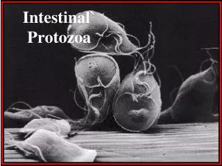
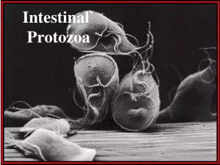
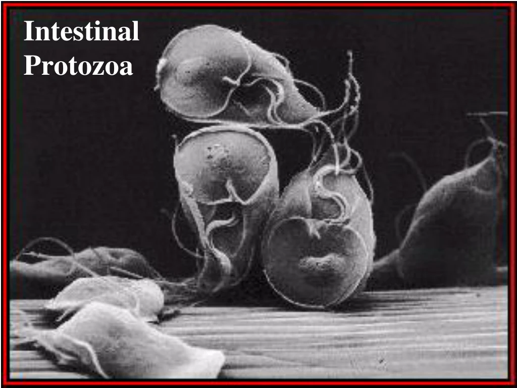
Intestinal Protozoa. CLASSIFICATION OF PARASITES. Giardia lamblia. Giardia trophozoites (SEM). Giardia trophozoites (light microscope). Flagellates : Giardia lamblia. Trophozoite. Cyct. Giardia cyst (light microscope) INFECTIVE STAGE.

E N D
Giardia lamblia Giardia trophozoites (SEM) Giardia trophozoites (light microscope)
Flagellates: Giardia lamblia Trophozoite Cyct
Giardia cyst (light microscope) INFECTIVE STAGE
Giardia lamblia: Life cycle
Giardia trophozoites in tissue section seen by duodenal aspirate
Giadriasis: Clinical Picture • The parasite mostly asymptomatic or can produce a wide range of gastrointestinal symptoms especially in children. • Symptomatic Infections: • Typical picture: Incubation period 1-2 wks followed by diarrhea, vomiting & flatulence for about 6 wks • Atypical: Severe diarrhea, malabsorption especially in children and cholecystitis
Giardiasis: Laboratory diagnosis • Stools examination • Microscopy for cysts or trophozoits • Detection of Giardia antigens in stools • Examination of duodenal contents: trophozoites
Giardiasis: Chemotherapy • Drug of choice: Metronidazole
ENTAMOEBA HISTOLYTICA… 500 million people are infected. 100,000 deaths per year. Worldwide distribution but is seen more often in tropical countries with poor sanitary conditions. It is a waterborne infection. There are 6 species of Entamoeba: E. histolytica E. dispar E. hartmanni E. coli E. gingivalis E. polecki
E. histolytica vs E. dispar Entamoebahistolytica:Amoebae that are pathogenic and invasive. E. dispar:The non-pathogenic, non invasive form. The 2 amoebae can’t be distinguish by microscopic observation.
Entamoeba histolytica Trophozoite: vegetative stage, must encyst to survive in the environment. It is a fragile structure. Cyst: infective stage. Resist the harsh conditions of the environment.
E. histolyticacyst E. histolyticatrophozoite
Entamoebahistolytica • Mode of infection (fecal-oral route) • Water, food • Flies can act as vector. • Can be sexually transmitted person to person contacts • Not a zoonosis
Entamoeba histolytica The infective dose can be as little as 1 cyst The incubation period can be from few days to few weeks depending on the infective dose. If the TROPHOZOITE is ingested it is disintegrates in the stomach without producing infection. Excystation occurs in the lower region of the small intestine and then production of 8 small amoebae which enter the large intestine and may: (1) invade the tissue, (2) live in the lumen of large intestine without invasion, or (3) encyst (become a cysts and pass in the stool). Only the Cysts can survive in the environment for weeks at appropriate temperature and humidity after excreted from stool of infected patients.
Entamoeba histolytica Intsetinal amoebiasis (Acute amoebic dysentry) : Trophozoite has the ability to hydrolyze host tissues with their active enzymes present on the surface membrane of the trophozoite, also trophozoite has the ability to ingest blood cells. The presenting symptom is diarrhoea which is accompanied by blood, mucus and sometimes tenesmus. As a complication, severe intestinal hemorrhage or rarely perforation may occur, lesions are found in cecum, appendix or colon. They may heal. If perforation of the colon occurs, this may lead to peritonitis that can lead to death. Amoeboma: Granulomatous mass obstructing the bowel.
PATHOLOGY: Intsetinal amoebiasis Complications
PATHOLOGY Intsetinal amoebiasis : Entamoeba histolytica
PATHOLOGY Intsetinal amoebiasis : Entamoeba histolytica
PATHOLOGY: Intsetinal amoebiasis : E. Histolytica in mucosa. Numerous trophozoites can be seen with ingested erythrocytes.
A 30-year-old male experienced diarrhea for two weeks with fever of 39° C, nausea, vomiting, malaise and right upper abdominal pain. Physical examination revealed hepatomegaly 6 cm below the right costal margin. CT scan showed a single hypodense mass in the rigth lobe of 7.8 x 5.2 cm, round, with well defined borders. Serology was positive for Enamoebahistolytica at 1/512. Amebic liver abscess was diagnosed.
THE CLINICAL OUTCOMES OF INFECTION WITH Entamoebahistolytica
Main Drugs for Treatment of Amoebiasis • Intestinal: • Asympromatic (cysts only): diloxanide furoate (Furamide) • Symptomatic(cysts and trophozoites): Metronidazole • Extra-intestinal: • Metronidazole
Laboratory Diagnosis of Amoebiasis • Intestinal: • Stools examination: • Wet mount (cysts and trophozoites) • Concentration methods (only cysts) • Serology (mainly for invasive infections): IHA , ELISA • Extra-intestinal: • Serology: IHA, ELISA • Microscopy of tissues or fluids
Cryptosporidium Diagnosis Cryptosporidium, safranin Cryptosporidium, acid-fast stain
Cryptosporidium Diagnosis Crypto-Gardia FAT
Cryptosporidiosis Treatment • Self-limited in immunocompetent patients • In AIDS patients: paromomycin