Download
1 / 33
330 likes | 336 Views
Chapter 6A The Peripheral Nervous System: Afferent. Divisionhttp://www.brainline.org/multimedia/interactive_brain/the_human_brain.html?gclid=CJroxvfmjaACFVth2godUkI6eA.

E N D
Chapter 6A The Peripheral Nervous System: Afferent Divisionhttp://www.brainline.org/multimedia/interactive_brain/the_human_brain.html?gclid=CJroxvfmjaACFVth2godUkI6eA
Describe the components (afferent and efferent) of the peripheral nervous system. This will be measured by lecture and laboratory exams.
Outline • Pathways, perceptions, sensations • Receptor Physiology • Receptors have differential sensitivities to various stimuli. • A stimulus alters the receptor’s permeability, leading to a graded receptor potential. • Receptor potentials may initiate action potentials in the afferent neuron. • Receptors may adapt slowly or rapidly to sustained stimulation. • Each somatosensory pathway is “labeled” according to modality and location. • Acuity is influenced by receptive field size and lateral inhibition. • PAIN • Stimulation of nociceptors elicits the perception of pain plus motivational and emotional responses. • The brain has a built-in analgesic system.
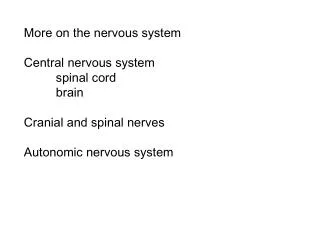
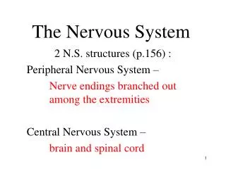
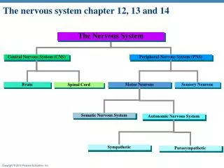
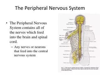
Peripheral Nervous System • Consists of nerve fibers that carry information between the CNS and other parts of the body • Afferent division • Sends information from internal and external environment to CNS • Visceral afferent • Incoming pathway for information from internal viscera (organs in body cavities) • Sensory afferent • Somatic (body sense) sensation • Sensation arising from body surface and proprioception • Special senses • Vision, hearing, taste, smell
Perception • Conscious interpretation of external world derived from sensory input • Why sensory input does not give true reality perception • Some information is not transduced • Some information is filtered out • Cerebral cortex further manipulates the data • Sensation vs. perception
What Do You Perceive? Proof !
Receptors • Structures at peripheral endings of afferent neurons • Detect stimuli (change detectable by the body) • Convert forms of energy into electrical signals (action potentials) • Process is called transduction
Types of Receptors • Photoreceptors • Responsive to visible wavelengths of light • Mechanoreceptors • Sensitive to mechanical energy • Thermoreceptors • Sensitive to heat and cold • Osmoreceptors • Detect changes in concentration of solutes in body fluids and resultant changes in osmotic activity • Chemoreceptors • Sensitive to specific chemicals • Include receptors for smell and taste and receptors that detect O2 and CO2 concentrations in blood and chemical content of digestive tract • Nociceptors • Pain receptors that are sensitive to tissue damage or distortion of tissue
Shaft of hair inside follicle Skin surface Epidermis Dermis Myelinated neuron Subcutaneous tissue Hair receptor: hair movement and very gentle touch Pacinian corpuscle: vibrations and deep pressure Ruffini endings: deep pressure Merkel’s disc: light, sustained touch Meissner’s corpuscle: light, fluttering touch Figure 6-5 p190
Muscle Receptors • Two types of muscle receptors. • Both are activated by muscle stretch, but monitor different types of information. • Muscle spindles monitors muscle length. • Golgi tendon organs detect changes in tension.
Muscle spindle (proprioceptor)regulates rate of change of length, And length Golgi tendon organ Type II sensory neuron Spinal cord Intrafusal muscle fibers Nuclear bag fiber Type lA sensory neuron Nuclear chain fiber Nuclei of muscle fibers Motor end plate Alpha motor neuron Extrafusal muscle fibers Gamma motor neuron Like pg. 289
Capsule Alpha motor neuron axon Intrafusal (spindle) muscle fibers Gamma motor neuron axon Contractile end portions of intrafusal fiber Afferent neuron axons Noncontractile central portion of intrafusal fiber Extrafusal (“ordinary”) muscle fibers Fig. 8-25a, p. 289
Uses For Perceived Information • Afferent input is essential for control of efferent output • Processing of sensory input by reticular activating system in brain stem is critical for cortical arousal and consciousness • Central processing of sensory information gives rise to our perceptions of the world around us • Selected information delivered to CNS may be stored for further reference • Sensory stimuli can have profound impact on our emotions
Receptors • May be • Specialized ending of an afferent neuron • Separate cell closely associated with peripheral ending of a neuron • Stimulus alters receptor’s permeability which leads to graded receptor potential • Usually causes nonselective opening of all small ion channels • This change in membrane permeability can lead to the influx of sodium ions. This produces receptor (generator) potentials. • The magnitude of the receptor potential represents the intensity of the stimulus. • A receptor potential of sufficient magnitude can produce an action potential. This action potential is propagated along an afferent fiber to the CNS.
Afferent terminals Rate of neurotransmitter release at afferent terminals +30 Afferent fiber potential (mV) Afferent fiber –70 Frequency of action potentials in afferent fiber Sensory receptor Receptor potential (mV) Rest Magnitude of receptor potential Stimulus strength Stimulus On On Off Off Time (sec) Stimulus strength Figure 6-3 p189
Receptors • May adapt slowly or rapidly to sustained stimulation • Types of receptors according to their speed of adaptation • Tonic receptors • Do not adapt at all or adapt slowly • Muscle stretch receptors, joint proprioceptors • Phasic receptors • Rapidly adapting receptors • Tactile receptors in skin
Phasic- Membrane potential drops More rapidly (intensity i.e pressure) Tonic -Takes longer for the membrane Voltage to drop (maintaining the signal i.e position) Fig. 6-5, p. 185
Somatosensory Pathways • Pathways conveying conscious somatic sensation • Consists of chains of neurons, or labeled lines, synaptically interconnected in particular sequence to accomplish processing of sensory information • First-order sensory neuron • Afferent neuron with its peripheral receptor that first detects stimulus • Second-order sensory neuron • Either in spinal cord or medulla • Synapses with third-order neuron • Third-order sensory neuron • Located in thalamus
Acuity • Refers to discriminative ability • Influenced by receptive field size and lateral inhibition
Lateral inhibition Fig. 6-7, p. 187
Pain • Primarily a protective mechanism meant to bring a conscious awareness that tissue damage is occurring or is about to occur • Storage of painful experiences in memory helps us avoid potentially harmful events in future • Sensation of pain is accompanied by motivated behavioral responses and emotional reactions • Subjective perception can be influenced by other past or present experiences
Cortex • Higher processing • Basal nuclei • Control of movement, inhibitory, negative • Thalamus • Relay and processing of sensory information • Awareness, a positive screening center for information • Hypothalamus • Hormone secretion, regulation of the internal environment • Cerebellum • Important in balance and in planning and executing voluntary movement • Brain Stem • Relay station (posture and equilibrium), cranial nerves, control centers, reticular integration, sleep control
Pain • Presence of prostaglandins (lower nociceptors threshold for activation) greatly enhances receptor response to noxious stimuli • Role of asprin • Nociceptors do not adapt to sustained or repetitive stimulation • Three categories of nociceptors • Mechanical nociceptors • Respond to mechanical damage such as cutting, crushing, or pinching • Thermal nociceptors • Respond to temperature extremes • Polymodal nociceptors • Respond equally to all kinds of damaging stimuli
Pain • Two best known pain neurotransmitters • Substance P • Activates ascending pathways that transmit nociceptive signals to higher levels for further processing • Glutamate • Major excitatory neurotransmitter • Brain has built in analgesic system • Suppresses transmission in pain pathways as they enter spinal cord • Depends on presence of opiate receptors • Endogenous opiates – endorphins, enkephalins, dynorphin
Higher processing of pain • Substance P • Different destinations • Cortex – localizes the pain • Thalamus- perception of pain • Reticular formation- increases alertness • Hypothalamus/limbic system- emotional and behavioral responses • Glutamate • AMPA receptors • Ap’s in the dorsal horn • NMDA receptors • Ca entry makes dorsal horn neuron more sensitive
(Localization of pain) Somatosensory cortex Higher brain (Perception of pain) Thalamus (Behavioral and emotional responses to pain) Hypothalamus; limbic system Brain stem Reticular formation ( Alertness) Noxious stimulus Spinal cord Afferent pain fiber Dorsal horn excitatory interneurons Substance P Nociceptor (a) Substance P pain pathway Figure 6-9a p195
Periaqueductal gray matter Reticular formation Medulla No perception of pain To thalamus Inhibitory interneuron in dorsal horn Endogenous opiate Noxious stimulus Opiate receptor Transmission of pain impulses to brain blocked Afferent pain fiber Dorsal horn excitatory interneurons Substance P Nociceptor (b) Analgesic pathway Figure 6-9b p195