Download
1 / 27
270 likes | 466 Views
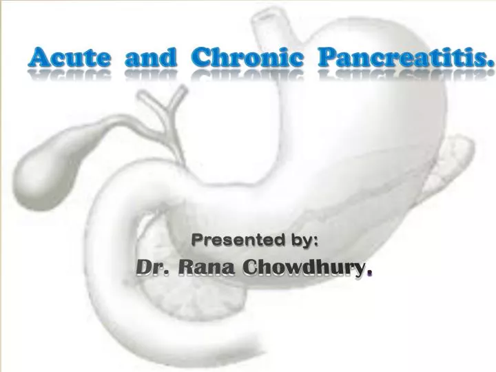
Acute and Chronic Pancreatitis. . Presented by: Dr. Rana Chowdhury . . Pancreas: A large gland behind the stomach that secretes digestive enzymes into the duodenum. . Acute Pancreatitis:

E N D
Acute and Chronic Pancreatitis. Presented by: Dr. Rana Chowdhury.
Pancreas: A large gland behind the stomach that secretes digestive enzymes into the duodenum.
Acute Pancreatitis: Acute abdominal pain usually associated with raised pancreatic enzyme level in the blood or urine as a result of inflammatory disease of pancreas.
Acute Pancreatitis: Pathogenesis: • Intracellular activation of trypsinogen to trypsin by numerous stimuli. • Activation, intestinal liberation and auto digestion of Pancreas by own enzymes. • Heredetery defect in chromosome – 7.
Acute Pancreatitis: causes: • Billiary stone (50 – 70%) . • Alcoholism (25%) . • Trauma (Surgical, Post ERCP, Blunt trauma etc.) • Drugs, Metabolic disorders. • Infections, like mumps, mycoplasma etc. • Idiopathic.
Acute Pancreatitis: symptoms: • Epigastric pain. • Nausea, Vomiting. • Fever.
Acute Pancreatitis: Signs: • Tachypnoea, Tachycardia, Hypotension. • Patient may be in shock. • Bleeding into Fascial plan produce - Gray Turner’s sign. - Culler’s sign. • Icteric in gall stone pancreatitis. • Small red tender nodule on leg.
Acute Pancreatitis: Signs on abdominal examination: • Abdomen distended due to paralytic ileus. • Tender epigastrium. • Muscle guard in epigastric region. • Bowel sound may be absent.
Acute Pancreatitis: Diagnosis • Serum Amylase (within 24 hours) • Urine Amylase (after 24 hours) • Ultrasonograph of whole abdomen • Serum lipase level • Serum Calcium level • Plain X ray abdomen: sentinel loop; colon cut off sign, renal halo (oedema around kidney) sign.
Acute Pancreatitis: management: Conservative: • Immediate hospitalization. • Bed rest. • Antispasmodic, analgesic. • Nothing per os, NG Suction, I/V fluid. • Non invasive monitoring. • Oral feeding after 7 days in mild & 14 days in severe case.
Acute Pancreatitis: management: Conservative: [contd.] • HDU/ ICU in severe cases, with narcotic for analgesia, invasive monitoring with ABG analysis, Catheterization, CVP etc. Surgery indicated in case of: • Diagnostic dilemma. • Acute haemorrhaegic pancreatitis. • Necrotizing pancreatitis.
Acute Pancreatitis: management: Surgery indicated in case of : [contd.] • Gall stone disease. • If patient does not respond to conservative treatment. ERCP: • Pancreatitis due stone impact in ampula of vater. • Abnormal LFT.
Acute Pancreatitis: prognosis: Ranson score:
Acute Pancreatitis: prognosis: Glasgow scale: On admission: Within 48 hours: Age > 55 years, Serum Calcium < 2 mmol/ L WBC Count > 15 X 109 / L Serum Albumin < 32 gm/ L Blood glucose > 10 mmol / L LDH > 600 units/ L Serum urea > 16 mmol / L AST/ ALT > 600 units/ L Arterial O2 saturation < 8 kPa
Acute Pancreatitis: complications: Local: • Acute fluid collection. • Sterile pancreatic necrosis. • Infective pancreatic necrosis. • Pancreatic abscess. • Pseudocyst. • Pancreatic ascitis. • Pleural effusion.
Acute Pancreatitis: complications: Systemic: • Shock, Arrythmia. • ARDS. • Renal failure. • DIC. • Hypocalcaemia, Hypoglycemia. • Visual disturbance, Confusion. • Subcutaneous fat necrosis.
Chronic Pancreatitis: Chronic pancreatitis is a chronic inflammatory disease in which there is irreversible progressive destruction of pancreatic tissue. • Male female ratio = 4 : 1 • Mean age of onset is above 40 years.
Chronic Pancreatitis: Etiology: • High alcohol consumption in 60-70% cases. • Pancreatic duct obstruction, resulting from stricture formation after • Trauma; • Acute pancreatitis; • Occlusion of duct by neoplasia or stone. • Congenital anomalies: pancreas divisum.
Chronic Pancreatitis: clinical features: • Pain: site of pain depends on the main focus of disease. • Nausea, vomiting. • Exocrine and endocrine pancreatic insufficiency. • Over and above, almost all complications of acute pancreatitis may be present in chronic pancreatitis.
Chronic Pancreatitis: Diagnosis: • Plain X ray abdomen may show pancreatic calcification. • CT scan or MRI can show outline of the gland, the main area of damage and possibilities of surgical correction. • MRCP will identify presence of Billiary obstruction & state of pancreatic duct. • ERCP is most elucidating for the duct anatomy.
Chronic Pancreatitis: Treatment: Medical treatment: • Low fat and high protein diet. • Pancreatic enzyme supplimentation. • Stop the patient from alcoholism & smoking. • Eliminate obstructive factors. • Escalate analgesia. • For intractable pain, consider CT guided coeliac axis block.
Chronic Pancreatitis: Prognosis: • Chronic pancreatitis is difficult to treat and often recurs. • Permanent exocrine or endocrine dysfunction. • Development of pancreatic cancer.
Bailey & Love’s – Short practice of surgery. • Current surgical diagnosis & treatment – Gerard M. Doherty. • Essential Surgical Practice – Sir Alfred Cuschieri.