Download
1 / 41
960 likes | 1.67k Views
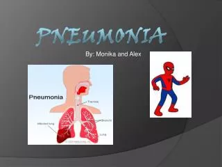
Pneumonia. Pneumonia. Definition: Acute infectious inflammation of the distal lung paranchyme (Distal to terminal bronchioles) with clinical and radiological signs of consolidation Pneumonitis: Noninfectious inflammation. Community Acquired Nosocomial (Hospital acquired)

E N D
Pneumonia • Definition: Acute infectious inflammation of the distal lung paranchyme (Distal to terminal bronchioles) with clinical and radiological signs of consolidation • Pneumonitis: Noninfectious inflammation
Community Acquired Nosocomial (Hospital acquired) Pneumonia in immuncompromised host Anatomic Lober Bronchopneumonia Interstitial pneumonia Etiologic Bacterial Viral Fungal Classifications
The microorganism reaches the lungs by: • Inhalation or aspiration • Hematogenious way • Direct invasion from the neighbouring tissues • The amount of the organism inoculated, the virulance factors and the immunity of the host are important factors
Most frequent • S. Pneumonia (50%) • H. İnfluenzae • Moraxella catarrhalis • Mycoplasma pneumonia • Chlamydia pneumonia • Legionella pneumophilia • Virus (10-20%) Atypical pn
Community acquired pneumonia • The symptoms of pneumonia are usually nonspecific but generaly include: • Fever (chills) • Cough • Sputum production (purulent) • Thoracic pain • Dyspnea
Typical pneumonia is characterised by abrubt onset high fever, chills, productive cough, thoracic pain, focal clinical signs, lobar or segmental radiographic findings, leukocytosis • Strep. Pneumonia • Rast colored (pink) sputum • Labial herpes lesions • Lober infiltration • H. influenzae
Different presentation • Confusion, tachypnea, hypotermia can be the presenting symptom in old age groups • Unusual presentation can be seen in immunocompromised patients
Atypical pneumonias are characterised by progressive onset, fever without chills, a cough without sputum, headache, myalgia, diffuse crackles, modest leukocytosis, interstitial infiltrates on chest radiographs. • Mycoplasma pneumonia • Legionella (bradicardia, hyponatremia) • Chlamydia
Physical examination • High fever, tachicardia, tachypnea, hypotension, confusion, drowsiness, altered mental status • Respiratory system: • Inspection: • Usually normal • Ortopnea • Cyanosis • Respiratory disstress • Palpation • İncreased Vibration thoracic (lober pneumonia) • Decreased hemithoracal movement
Percution • Normal sonority • Dullness (Matite) • Oscultation • End inspiratory fine crackles • Local diminished breath sounds • Bronchial voice
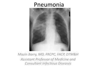
Diagnosis • History and symptoms • Physical examination • PA Chest x-ray • Microbiologic examination • Routine laboratory tests • CBC,ESR,CRP,Hepatic enzymes,Renal functions • Blood gas
Consolidation Lobar or patchy nonhomogenious infiltrations Air bronchogram Round opacity Fine reticular density Complications Pleural effusion Cavitation Abscess Pneumatocell Pneumothorax PA Chest x-ray
Microbiologic examination(identification of the causative pathogen) • The causative pathogen can not be isolated in 30-50% of CAP (Not always necessary) • Sputum • Gram Staining (more specific than culture but less sensitive) In microscopic examination sputum shoud show <10 epithelial cell , and >25 PNL • Culture (Staining and culture shoud be consistent) • Blood culture (Hospitalised patients) • Pleural fluid
Serology (Urine, sputum or blood: pneumococcal antigen, urine: Legionella antigen, DFA, 4 fold increase in specific antibody titers (cold agglutinins) between acute and covalescent period • İnvasive techniques (Shoud be performed in severe cases and immunocompromised patients) (FOB, BAL, Protected-brush, TBB, PCFNA)
Approach to the patient • Is it pneumonia? • How severe is the illness? • Outpatient treatment? • Hospitalization? • Intensive care? • Risk factors • Severe condition
Age>65 Comorbid illness Alcoholism Aspiration? Recurrent pneumonia <1year Mental problems Spleenectomy Malnutrition Social problems CS use >10 mg prednisolone for 3 months Immunosuppressive treatment Pneumonia following influensa Risk Factors
Signs of Severe condition • Respiratory rate >30/min • BP <90/60 mmHg • Fever>38,3 C • Extrapulmonary disease (menegitis, artritis, myocarditis etc) • WBC <4000 or >30000 / mm3 • Htc <30% or Hb<9 gr/dl • ABG PaO2<60 mmHg PCO2>50 mmHg • BUN >20 mg/dl • Multilober infiltration, cavity, effusion, rapid progression • Sepsis or multisystem disfunction
CURB-65 Score • Confusion • Urea>42.8 mg/dl; BUN>20 mg/dl • RR>30/min • BP<90/60 mmHg • Age>65 Predicting mortality, each is 1 point. A score >2 points Hospitalization
Probable microorganism S. pneumoniae M. pneumoniae Chlamydia pneumoniae H. influensa Virus Other Empirical Treatment Amoksisilin 1gr/8hr Macrolid (Klaritromycine, azitromycine) or Doksisiklin (According to clinical signs (atypical?) or allergic conditions) Group I A Outpatient treatment Risk factor(-) Severe condit ion (-)
Probable microorganism S. pneumoniae M. pneumoniae Chlamydia pneumoniae Mikst infeksiyon H. İnfluensa Enterik Gr (-) Virus Other Empirical Treatment 2-3. line sephalosporin (nonpseudomonal) Beta-laktamase inhibiting aminopenisilin ± Macrolid veya Doksisiklin (In case of intolerability, allergy only florokinolon Moksifloksasin, Levofloksasin) Risk factor (+) Severe condition (-) Group I B Send to hospital Outpatient treatment
Probable microorganism S. pneumoniae H. İnfluensa M. pneumoniae Chlamydia pneumoniae Mixed infection Aerob Gr (-) Anaerobic Legionella Virus Group 2 Severe condition (+)and/or Risk factor (+) Hospitalized Empirical treatment: 3. line nonpseudomonal sephalosporin or beta laktamase inhibiting aminopenisilin + Macrolid /Doksisiklin Or Florokinolon alone
RR>30 PaO2/FiO2 ≤250 Confusion/ disorientation BP<90/60 mm Hg Urine <20 ml/st,(BUN>20 mg/dl) ARF WBC<4000/mm3 PLT<100 000/mm3 Temp<360C Bilateral, multilober infiltration or progression >50% in 48 hrs Hypotension that needs heavy iv support Indications for mechanical ventilation Septic shock (need for vasopressor drugs) Intensive Care Indications
Probable microorganism S. Pneumoniae Legionella H. İnfluensa Enteral Gr (-) S aureus M pneumonia Virus Other Probable microorganism P aureginosa Grup A pathogens B Pseudomonas risk(+) Group 3 APseudomonas risk(-) Intensive care tr. indication (+) Empirical treatment: 3. Line nonpseudomonal sephalosporin or beta laktamase inhibiting aminopenisilin + Macrolid or Florokinolon (Add rifampicin if documented legionella+) Antipseudomonal betalaktam + Ciprofloksasin/ofloksasin or aminoglikozid + Macrolid (in non Kinolon combined group)
Risk for Pseudomonas • Underlying lung disease (Bronchiectasis, C. Fibrozis, severe COPD) • Steroid (>10 mg/gün) • Antibiotic use (>7 days in the previous month) • Malnutrition
Certain risk-pathogen relations • Gr (-) enteral bacillei • Nursing home residency • Concomitant CVS disease • Multipl concomitant disease • Recent antibiotic use • 3rd generation cephalosporines, fluorokinolones (3-4 weeks) • Antipseudomonal penicillines, ceftazidime +aminoglicoside for pseudomonas • Anaerob bacteria • Poor oral hygen • Probability of aspiration (alcoholism, epileptic atack, gingivitis, esophageal obstruction • iv drug abuse • Obstructive bronchial pathologies • Fusobacterium, bacteroides, peptostreptococcus, actinomyces • Sputum with bad smell, • Betalactamase inh aminopenicilins, penicillin G, clindamycine, metranidazole, ornidazole (4-6 weeks if necrosis is present)
Legionella pneumophila • Age >65 • Malignancy • COPD • Steroid treat. • Smoking • Recent travel (hotel) • Water supply system reconstruction • Macrolide (21 days) • Rifampicine, kinolones • S. Aureus (rapid progression, cavitation, severe illness) • Concurrent influensa epidemic, • Nursing home resident • Iv drug abuse • Vancomycine, Teikoplanin (min 3 weeks, 6 weeks if abscess is formed) • C. psittachi • Recent bird contact • At risk occupation
Follow up • Parenteral to oral tr. shift: • Afebril period of 24 hours • Decreased neutrophylia • Clinical stability • Decreased CRP>50% • Treatment response: • Evaluated in 72 hours unless a resistant bacillei is shown or clinical deteoriation • Radiologic control in 7-10 days • Radiologic complete resolution may take 4 weeks, can be longer in elderly, alcoholics, COPD patients • Treatment period • Pnomococ 7-10 days • Mycoplasma, Clamydia 10-14 days • Legionella 14-21days • Unknown 2-3 weeks
Prevention • Influensa vaccine • Pneumococ vaccine • General hygene • Staff education (hand washing, glowes) • Avoid invasive procedures if possible • Sucralphate for gastric prophylaxis • Enteral feeding as much as possible • Avoid narcotics • Early mobilization • Early discharge from IC or hospital CAP Nasocomial
Certain Definitions • Recurrent pneumonia: A second pneumonia that occurs after the complete healing of a first attack (>1 month). At least 2 times a year. • Late resolution: A pneumonia that resolves <50% in 2 weeks or incomplete regression in 4 weeks • Nasocomial Pneumonia: Pneumonia seen after 48 hours of hospitalization or within 48 hours after being discharged from hospital
Age COPD Alcoholism Smoking D mellitus Malignancy Renal or cardiac failure CS use S pneumonia Legionella Viral H influensae Risk factors for late resolution:
Complications of pneumonia • Pleural effusion (parapneumonic) • Emphyema • Bronchopleural fistule • Mediastinitis, pericarditis, chest wall infection • Necrosis, cavitation • Pneumatocel • Pneumothorax • ARDS • Fibrosis • Bronchiectasis • Late resolution or recurrens