Download
1 / 55
550 likes | 559 Views
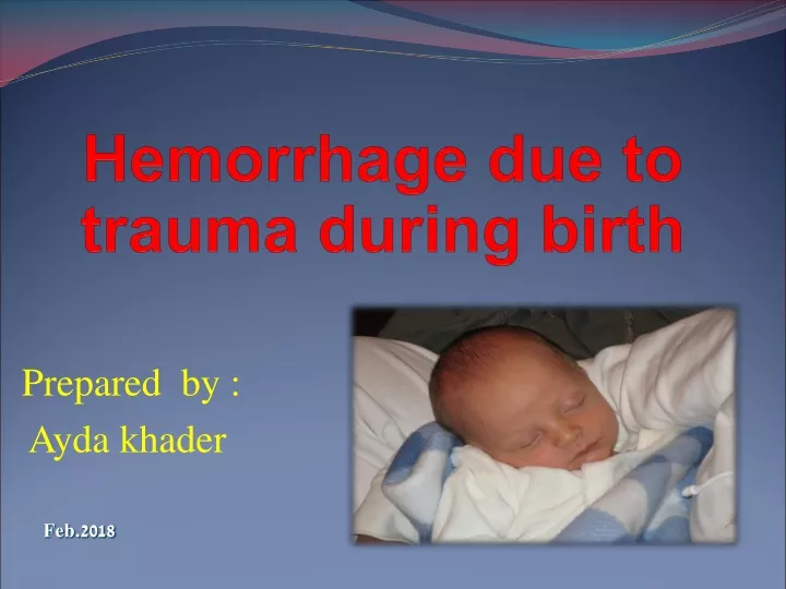
Hemorrhage due to trauma during birth. Prepared by : Ayda khader. Feb.2018. Outline. major types of neonatal haemorrhage due to trauma Haemorrhage due to distruption in blood flow Neonatal convulsions specific interventions with parent. Blood volume is approximately

E N D
Hemorrhage due to trauma during birth Prepared by : Ayda khader Feb.2018
Outline • major types of neonatal haemorrhage due to trauma • Haemorrhage due to distruption in blood flow • Neonatal convulsions • specific interventions with parent
Blood volume is approximately • 80–100 ml/kg in the term baby • pre-term baby 90–105 mL/kg therefore even a small haemorrhage can be potentially fatal.
Haemorrhages can be due to: • trauma • disruptions in blood flow or can be related to: • coagulopathies • other causes.
Hemorrhages types: • Extra cranial • Caput succedaneum • Cephalhematoma • Subgaleal Hemorrhage • Intracranial • Epidural • Subdural • Subarachnoid
A cephalhaematoma is an effusion of blood under the periosteum that covers the skull bon
I -Haemorrage due to trauma Cephalhematoma • is a subperiosteal collection of blood between the skull and the periosteum • It may be unilateral or bilateral, • appears as firm, does not pit on pressure ,swelling on theside of the head. • A cephalhaematoma never extends beyond the edges of the bone • does not cross suture lines, no overlying skin discoloration • is not present at birth; the swelling appears after 12 hrs, grows larger over subsequent days
Occure due to with friction between the fetal skull and maternal pelvic bones, such as in cephalopelvic disproportion or precipitate labour, the periosteum is torn from the bone • Occurs after prolonged labor and instrumentation, vacuum-assisted births. • subsequent days and can persist for weeks., does not cross a suture and is fixed • No treatment is necessary and the swelling subsides when the blood is reabsorbed. • Haemolysis of the extravasated blood may result in hyperbilirubinaemia.
Subgaleal Hemorrhage • SubaponeuroticHemorrhage also called • Occurs between periosteum and epicranialaponeurosis • may occur with any type of birth but is more often associated with vacuum-assisted births • The swelling is present at birth, increases in size • This a firm, fluctuant mass ,The scalp is movable rather than fixed. • The swelling can cross sutures and extend into the subcutaneous tissue of neck and eyelids. • The baby experiences pain with head movement or handling of the swelling.
Some subaponeurotic haemorrhages are small but there is a risk of excessive haemorrhage and severe shock. • This emergency situation requires immediate medical assistance; stabilization and full supportive care, including blood transfusion. Subaponeurotic haemorrhage is associated with a mortality rate of 25% • the blood is reabsorbed and the swelling and bruising resolve over 2–3 weeks • Hyperbilirubinaemia complicates recovery
Anatomy of an Epidural Bleed • Three membranes (the meninges) • envelop the brain and spinal cord: pia, arachnoid, and dura
The epidural space: • the space between the dura (the outermost membrane covering the brain and spinal cord) and skull, or the bony vertebrae that form the spinal canal. In the spine, the epidural space contains lymphatics, small arteries, and the epidural venous plexus. The subdural space: • The space between the dura mater and the arachnoid mater, this is a potential space in both the skull and the spine. The subarachnoid space: • between the arachnoid mater and the pia mater, this space contains the cerebrospinal fluid
Epidural haemorrhage • Rare • Usually associated with skull fractures • Irritability, lethargy, and seizures progress to signs of increased ICP • Diagnosed by CT
Intracranial Hemorrhages: • Subdural haemorrhage • May be due to rupture of the straight sinus, vein of Galen, transverse sinus, inferior sagittal sinus, or superficial bridging vessels • Predisposing factors include rapid, abnormal or excessive moulding, such as in precipitate labour or rapid birth, malpositions, malpresentations, cephalopelvic disproportion, or undue compression during forceps manoeuvres • Subdural haemorrhage may be fatal.
A baby with a small haemorrhage may demonstrate no signs and resolution is spontaneous • if blood continues to leak, the signs develop over several days. As blood accumulates, there is cerebral irritation, cerebral oedema and raised intracranial pressure • baby is likely to vomit, be non-responsive, and have a bulging anterior fontanelle, abnormal eye movements, apnoea, bradycardia and convulsions
Diagnosis is confirmed by cranial USS ,computerized tomography (CT),magnatic resonance imaging (MRI) • Supportive treatment focuses on replacing blood volume and controlling the consequences of asphyxia and raised intracranial pressure • Surgery to relive pressure or shunt placement
II-Intracranial haemorrage due to disruption in blood flow • Subarachnoid haemorrhage • A primary subarachnoid haemorrhageinvolves bleeding directly into the subarachnoid space • Pre-term babies who suffer hypoxia at birth resulting in disruption of cerebral blood flow, and term babies who suffer traumatic births are vulnerable • A secondary haemorrhage involves leakage of blood into the subarachnoid space from an intraventricular haemorrhage
often asymptomatic • baby may have generalized convulsions from the second day of life, pre-term babies may have apnoeic episodes • Subarachnoid haemorrhage is difficult to see on USS, although computerized tomography (CT) scanning can demonstrate the haemorrhage. • .If a lumbar puncture is performed, the CSF will be uniformly bloodstained.
Management includes replacement of blood volume • control of the consequences of asphyxia and convulsions. • The condition is usually self-limiting
Intraventricular haemorrhage(IVH) • Also called Germinal matrix haemorrhage (GMH) and intraparenchymal lesions (IPL) • primarily affect babies of <32 weeks' gestation and those weighing <1500 g • The incidence and severity of these haemorrhages /lesions are inversely correlated with gestational age • Has 4 grade
Grades • Based on the extension of the hemorrhage • Ventricular measurement • Mild dilation: 3-10 mm • Moderate dilation: 11-14 mm • Large dilation: greater than 14mm
A grade I • haemorrhage into the germinal matrix, is a periventricular or sub parenchymal haemorrhage • Without ventricular enlargement A grade II • Extension of the haemorrhage into the lateral ventricle • Minimal ventricular enlargement
A grade III • If a grade II haemorrhage is complicated by blockage to the outflow of CSF, post-haemorrhagic hydrocephalus develops and the ventricles dilate; • Moderate or large ventricular enlargement A grade IV • haemorrhage may extend into the cerebral tissue, giving rise to a periventricular/parenchymal haemorrhage • Intraparenchymal hemorrhage
factors may affect cerebral haemodynamics resulting in GMH/IVH/IPL. Early factors include obstetric haemorrhage, lack of antenatal steroids, birth outside a regional unit, chorioamnionitis, low one minute Apgar score, bruising at birth and low umbilical artery pH. Later risk factors include acidosis, hypotension, hypertension, respiratory distress syndrome (RDS) requiring mechanical ventilation, ‘fighting the ventilator’, apnoea, rapid volume expansion, rapid administration of sodium bicarbonate, pneumothorax and patent ductus arteriosus . Also implicated are excessive handling, exposure to light and noise, lateral flexion of the baby's head and crying.
Clinical picture • Approximately 50% of GMHs are small, have a ‘silent’ onset, and are detectable only on USS. • If the haemorrhage is larger or extends, the clinical features may gradually appear and worsen, • including apnoeic episodes that become more frequent and severe, bradycardia, pallor, falling packed cell volume, tense anterior fontanelle, metabolic acidosis and convulsions. • The baby may be limp or unresponsive. If the haemorrhage is large and sudden in onset, apnoea and circulatory collapse may present
Care of at-risk babies is focused on prevention • If complications develop, such as pneumothorax or PDA, these should be quickly detected and effectively treated • The baby's developmental needs should be met, particularly in relation to supportive flexed positioning, reduction in bright lighting, a quiet, undisturbed environment and appropriate interaction with parents and others • The outcome depends on the nature of the lesion. The neurological prognosis for babies with a GMH or a small IVH is usually good
IVH associated with ventricular dilatation may resolve spontaneously with no long-term consequences. • a large IVH and ventricular dilatation, the accumulating CSF may require temporary drainage using ventricular taps or external ventricular drainage • Some babies may require permanent CSF drainage via a ‘shunt’ • The prognosis for these babies is less good. • Approximately 50% of babies with a massive IVH die.
Intracerebral hemorrhage • cerebral hemorrhage is uncommon in all newborns, An intracerebral hemorrhage may occur rarely as a primary event related to rupture of an arteriovenous malformation or aneurysm, from a coagulation disturbance (e.g., hemophilia, thrombocytopenia), or from an unknown cause.
More commonly, cerebral intraparenchymal hemorrhage (IPH) occurs as a secondary event, such as hemorrhage into a region of hypoxic-ischemic brain injury • Intracerebellar hemorrhage occurs more commonly in preterm than term newborns and may be missed by routine CUS.
The presentation differs depending on the size and location of the IPH • In the preterm infant, IPH is often clinically silent unless the hemorrhage is quite large • In the term infant, intracerebral hemorrhage typically presents with focal neurologic signs such as seizures, asymmetry of tone/movements along with irritability or depressed level of consciousness. • Diagnosis MRI is the best imaging modality for IPH, but CT may be used in the preterm infant or when a rapid bedside imaging study is necessary.
Management • Small hemorrhages require only symptomatic treatment and support. • Large IPH with severe neurologic compromise should prompt neurosurgical intervention
Haemorrhage related to coagulopathies • These haemorrhages occur due to disruption of the baby's blood-clotting abilities. Vitamin K deficiency bleeding • Vitamin K deficiency bleeding (VKDB) may occur up to 6 months of age • more commonly occurs between birth and 8 weeks of life. • Several proteins, factor II (prothrombin), factor VII (proconvertin), factor IX (plasma thromboplastin component), factor X (thrombokinase) and proteins C and S, require vitamin K for their conversion to active clotting factors.
A deficiency of vitamin K, as in VKDB, leads to a deficiency of these clotting factors and resultant bleeding. • Neonatal hepatic immaturity impairs the synthesis of these factors and limits the effectiveness of vitamin K. • Inefficient transplacental transfer of vitamin K also may be a contributing factor. • Delayed colonization of the gut by bacteria (which become an endogenous source of vitamin K) is another factor that occurs with delayed feeding, breastfeeding, vomiting, severe diarrhea, and the use of antibiotics
Presentation Classic vitamin K deficiency presents in the first week of life (except first 24 hrs) with • epistaxis, purpura • large cephalohematomas, intracranial hemorrhage • melena, GI, GU bleeding • bleeding from the umbilical stump, injection sites, and after circumcision. • Bleeding occurs most commonly from the second to seventh days of life in healthy, solely breastfed, and often term infants who have not received prophylactic vitamin K.
It occurs rarely in formula-fed infants because of the supplemental vitamin K in those products. • When prophylactic vitamin K is not administered, the frequency of clinical bleeding averages 1.5%. • much more common than the early or late forms of the disease. Early vitamin K deficiency • severe bleeding within the first 24 hours of life • linked to maternal use of medications (most often anticonvulsants (phenobarb, phenytoin), coumadin, also anti-Tb meds) that interfere with vitamin K stores or function.
Classical VKDB (1-7 days) developing with those who are preterm or low birth weigth they are more likely to spontaneous bleed or have invasive intervention resulting in bleeding that cannot be controlled Distruption to the colonozation of the bowel
Late vitamin K deficiency (1-6 monthes )peak before 8week is linked to a compromised supply of vitamin K or malabsorption, as seen with • exclusive breast feeding without vit K • diarrhea • cystic fibrosis of the pancreas • hepatitis • biliary atresia • celiac disease • may cause: CNS and other life-threatening bleeding
Management • all pre-term babies, all ‘at risk’ term babies, and healthy term babies whose parents choose, an intramuscular injection of vitamin K1 should be administered within one hour of birth to give a transient peak serum concentration, reducing the risk of early VKDB • oral prophylaxis regimen should be instituted. • Oral vitamin K1 2.0 mg is given on the first and seventh days of life. • administration of vitamin K1 to lactating women increased the levels of vitamin K1 in the maternal serum and breastmilk • All parents should be given the opportunity to discuss vitamin K1 prophylaxis during pregnancy,
Thrombocytopenia • defined as a platelet count of <150 000/μLand severe thrombocytopenia is a platelet count of <50 000/μL • results from a decreased rate of formation of platelets or an increased rate of consumption. • In the general neonatal population, thrombocytopenia is rare
Risk of developing thrombocytopenia include : • a severe congenital or acquired infection (e.g. syphilis, cytomegalovirus, rubella, toxoplasmosis, bacterial infection) • isoimmunethrombocytopenia • inherited thrombocytopenia OR whose mother: • has idiopathic thrombocytopenia, purpura, systemic lupus erythematosus or thyrotoxicosis • takes thiazide diuretics.
Fetal thrombocytopenia, due to congenital infection or an inherited condition, may be monitored during pregnancy to determine the need for maternal immunoglobulin administration and/or prenatal intrauterine platelet transfusions • Postnatally, early onset thrombocytopenia is mild and due to a lack of platelet production associated with placental insufficiency • A petechial rash appears soon after birth, presenting in a mild case • a severe case there is widespread and serious haemorrhage from multiple sites. Intracranial haemorrhage may be fatal
Diagnosis • based on history, clinical examination and a reduced platelet count. • Mild early onset cases are usually self-limiting, and require no treatment • intravenous immunoglobulin administration may help • In severe cases, transfusions of platelet concentrate are required
Disseminated intravascular coagulation (DIC) • an acquired coagulation disorder associated with the release of thromboplastin from damaged tissue, stimulating abnormal coagulation and fibrinolysis with widespread deposition of fibrin in the microcirculation and excessive consumption of clotting factors and platelets • DIC is secondary to primary conditions • Maternal causes include pre-eclampsia, eclampsia and placental abruption.
Fetal causes include severe fetal distress, the presence of a dead twin in the uterus and traumatic birth. • Neonatal causes include conditions resulting in hypoxia and acidosis, severe infections, hypothermia, hypotension and thrombocytopenia • The diagnosis is made from clinical signs and laboratory findings that show a low platelet count, low fibrinogen level, distorted and fragmented red blood cells, low haemoglobin and raised fibrin degradation products (FDPs) with a prolonged PT and PTT