Download
1 / 25
270 likes | 347 Views
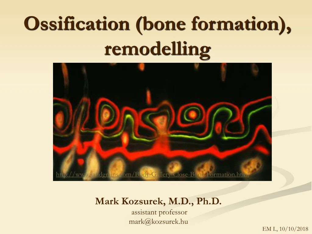
Ossification ( bone formation ), remodelling. http://www.sandgrains.com/Body-Gallery-Close-Bone-Formation.html. Mark Kozsurek, M.D., Ph.D. assistant professor mark@kozsurek.hu. EM I., 10/10/2018. Cells of the bony tissue. Osteons are only found in the compact bone!.

E N D
Ossification (boneformation), remodelling http://www.sandgrains.com/Body-Gallery-Close-Bone-Formation.html Mark Kozsurek, M.D., Ph.D. assistant professor mark@kozsurek.hu EM I., 10/10/2018
Cells of thebonytissue Osteons are only found in the compact bone! Periosteum contains mesenchymal cells!
1. Osteocyte: The most common cell type in mature bone. Rest in the lacunae of the bone. Adjacent osteocytes are interconnected by delicate processes. They arise from osteoblast, in fact they are the inactive forms of them.
2. Osteoblast: Differentiate from mesenchymal stem cells and are charachteristic for developing and regenerating bones as they are active and synthesize the organic components of the bony tissue called osteoid. Later they also contribute to mineralization and hydroxyapatite deposition.As they are synthesizing cells have an extended rER with ribosomes and due to these are basophylic. Mesenchymal stem cells in culture
3. Osteoclast: Osteoclastsaregeneratedbythefusion of severalmacrophages and arecrucialintheresorption of thebonytissue.
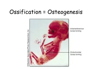
Boneformation • Primaryangiogenicossification of Krompecher: bone is synthesizedde novobyosteoblastsdifferentiatedfrommesenchymalcellsfound in theadventitia of smallvessels – it is commonduringremodelling of thebone. • Secondaryossifications • Bonereplaces a formertissuetype: • Intramembranousossification: osteoblastsdifferentiatefrommesenchymalcells and replacethepreexistingconnectivetissue. Somebones of theskull (e.g. thecalvary) and theclavicledevelopsbythistype of boneformation. • Endochondralossification: firstthehyalinecartilage is eroded, removedthenreplacedbyosteoidproducingosteoblasts. The wholeprocess is betterseen in tubularbones, butbelowtheclavicleallthebones of the human body formthisway.
Within a poorlydifferentiatedconnectivetissuesomemesenchymalcellstransformintoosteoblasts and formossificationcentres. • Osteoblastssynthesize and releaseosteoid, theorganiccomponent of thebonewhichcalcifiessoon. Trappedosteoblaststurnintoinactiveosteocytes. Onthe free surfaces of bonytrabeculaenewosteoblastsderivedfromsurroundingmesenchymalcellsaccumulate. Followingdeposition of newosteoidtheybecometrappedinactiveosteocytes. And thecyclegoeson… • Bone is formed in a random mannerfirstresulting in an irregularnetwork (wovenbone). Condensation of thevascularizedmesenchymeonthesurface of thedevelopingbonegivestheperiosteum. • Undertheperiosteumwovenbone is replacedbymaturelamellarbone (Haversiansystem) and compactplatesarecreated. Insidetheirregulartrabeculaepersist and thehiglyvascularizedtissuefillingthecavities of thespongybonediffenetiateintoredbonemarrow.
B M M B OCL M B OCL M OB OC Developingcalvary. Bonytrabeculae (B) encloseinactiveosteocytes (OC) and possessosteoid-synthesizingosteoblasts (OB) mainlyontheirexternalsurface and osteoclasts (OCL) ontheoppositesides. Osteoblastdifferentiatefrommesenchymalcells. Osteoclastsaregeneratedbythefusion of severalmacrophages and arecrucialintheresorption of theboneytissue.
OB B OC OCL OCL M
B) Endochondralossification Apoptotic chondrocytes are removed by chondroclasts and osteoblasts differentiating from mesenchymal cells sattle down on the surfaces of preserved mineralized inerterritorial matrix of cartilage.
1. 2. EPIPHYSEAL PLATE: • zone of resting cartilage • zone of proliferation • zone of degenaration and matrixcalcification • zone of mesenchymalinvasion 3. 4.
During the fetal period the hyalin cartilage model of the tubular bones appear. Perichondrium has mesenchymal cells some of which differentiate into osteoblasts, thus, the perichondrium is slowly replaced by periosteum. Periosteum and increasing number of osteoblasts arising from that form a bony sheath which is against the diffusion of nutrients and oxygen: hyaline cartilage degenerates and the interteritorial matrix calcifies. Apoptotic chondrocytes are removed and osteoblasts aggregate on the surfaces of trabeculae constituted by the persisting mineralized matrix. Osteoid sythesis begins and primary ossification centers develop still in the fetal period. Due to the invasion of small vessels into the epiphyseal cartilages and appearance of mesenchymal cells differentiating into bone-producing osteoblasts secondary ossification centers appear during childhood. Primary and secondary ossification centers are getting isolated by the epiphyseal cartilage/plate. This disk-like structure with its proliferatating chondrocytes is the source of longitudinal growth of these bones. Longitudinal growth is only possible while epiphyseal plates are present. They will slowly disappear by puberty due to the increasing concentration of sexual hormons.
Secondaryossificationcenters, epiphyseal plates 4 ½ years 11 years adult
zone of proliferation zone of degeneration and matrixcalcification (swollencellswithpycnoticnuclei, darkerinterterritorialsubstance)
chondroclast zone of degeneration and matrixcalcification redbonemarrow
bonytrabecula (spicule) osteocytesinside, osteoblastsonitssurface
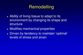
Boneremodelling A lifelong process where maturebone is resorbed and a newbone is synthesized. Remodellingmightalso be consideredas an adaptationtochangingdemands of mechanical loadings. In the first year of life, almost 100% of the skeleton is replaced. In adults, remodeling proceeds at about 10% per year. 1. Onthesurface of thetrabeculae of thespongybone Blood vessels are important as they are the sources of monocytes differentiating into osteoclasts.
2. In theHaversiansystems of thecompactbone mesenchymal cells Osteoclasts (from the monocyte-macrophage system) distroy concentrically arranged bony lamellae of the osteon and the resorption cavity is formed. Mesenchymal cells of the adventitia of small vessels differentiate into osteoprogenitor cells and osteoblasts which bild a new Haversian system.
blood vessel mesenchymal cells osteoblasts osteoclasts osteocytes in the old bone
puberty osteoblast osteoclast activity approx. 30-35 years
Hormonalcontrol of bonemetabolism plus: growth hormone, sexual steroids, thyroxin, vitamin-D, etc.
Paget’s disease Uncontrolled bone resorption and synthesis resulting irregularities.