Download
1 / 32
320 likes | 324 Views
This research aims to characterize the structural changes in chloroplasts and their relationship with the vacuole during leaf senescence in Arabidopsis. The study will utilize transmission electron microscopy (TEM) and confocal microscopy techniques to visualize the subcellular structures and assess the relevance of the vacuole in chloroplast senescence.

E N D
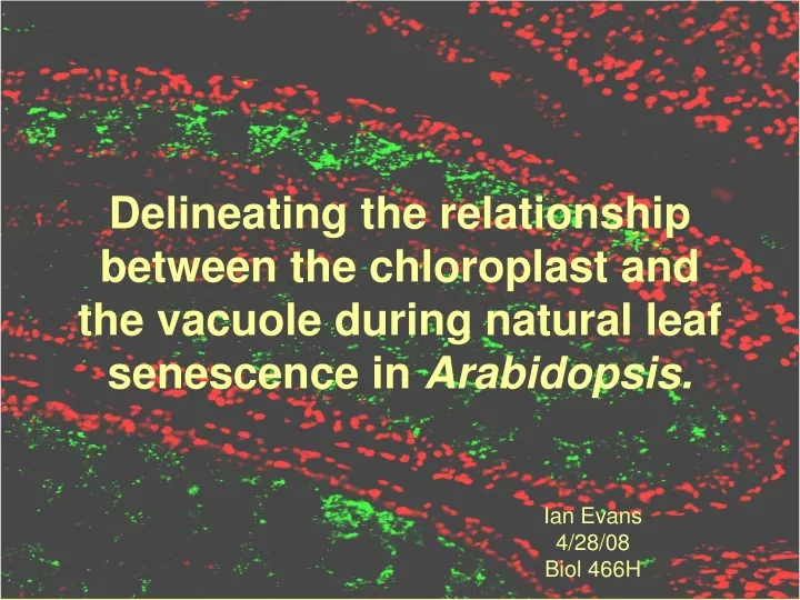
Delineating the relationship between the chloroplast and the vacuole during natural leaf senescence in Arabidopsis. Ian Evans 4/28/08 Biol 466H
Defining Senescence in Plants • Foliar senescence is the final stage of leaf development in which nutrients are remobilized to younger tissues
Lim et al. (2007) Annual Review of Plant Biology 58:122. Senescence is tightly regulated
Chloroplast (Cp) Senescence Chlorophyll degradation is well characterized, but degradation of the LHC proteins that bind Chl is not well understood. Hörtensteiner, S (2006) Annual Review of Plant Biology 57:55.
Evidence for Vacuolar Degradation • Endopetidases are present in vacuole and their activity is upregulated in senescence • TEMs show Cps in the center of the cell during senescence but not during normal growth
Wittenbach et al. Plant Physiol. 1982 69:98 Wheat Mesophyll Cell
Minamikawa et al. Protoplasma. 2001 218:144 Bean Mesophyll Cell
Evidence for Internal Degradation • Upregulation of Cp-localized proteases • Initial Chl catabolism occurs in the stroma • Early literature shows internal changes in Cps
Barton et al. Planta. 1966 71:314 Bean Mesophyll Cell senescing Cps
Freeman et al. 1978 Protoplasma 94:221 Citrus Mesophyl Cell senescing Cp
Aims of my research • Aim 1: Characterize the ultrastructure of the Cp in senescence • Aim 2: Define the relationship between the Cp and the vacuole in senescence
Rationale for Approach to Aim 1 • TEM will allow subcellular structure to be analyzed within senescent cells • Morphological changes in Cp will be evident
Zoning an Arabidopsis leaf Green Zone 3 Zone 2 Zone 1
Chlorophyll levels • Chl levels decrease as leaf senescence progresses
Through zoning system Rubisco is serially degraded Zone 1 shows absence of intact Rubisco with increased non-specific Ab binding Marker Green Zone 3 Zone 2 Zone 1 Rubisco levels 55kD
Ultrastructure of Leaves • Use Transmission Electron Microscope (TEM) to visualize subcellular structure within each zone
Healthy Green Cp Green 15,000X
Plastoglobuli formation Zone 3 15,000X
Separation of Thylakoids Zone 2 15,000X
Dismantling of Thylakoid System Zone 1 20,000X
Circularization and loss of distinguishable grana Zone 1 40,000X
Late Stage Separation from Plasma Membrane Zone 1 40,000X
Aim 1 Conclusions • While Chl and Rubisco are degraded, Cps remain intact along the plasma membrane • Ultrastructural changes include separation of thylakoids, formation of plastoglobuli, and circularization of Cps.
Rationale for Approach to Aim 2 • TEM is impractical even with serial sectioning or immunogold staining • Newer techniques are looked upon favorably • Confocal has less artifacts and preparation problems
Structure of the Vacuole • The tonoplast surrounds the central vacuole in plant cells
Confocal Microscopy • To visualize the tonoplast - use a 35S-GFP::d-TIP line. To visualize the chloroplast - use autofluorescence.
Avila et al. 2003. Plant Phys. 133:1674 • GFP lines treated with EMS to induce single nt mutations • Above, tvs mutant’s vacuoles transected by transvacuolar strands • To the right, bub mutants have many small vacuoles
Eggink, et al. 2004. BMC Plant Biology, 4:5 http://icecube.berkeley.edu/~bramall/work/astrobiology/images/chlorophyllspectra.jpg
What would results look like? During normal growth: During senescence:
Conclusions and Significance • Degradation of chloroplast will be characterized in Arabidopsis • Relevance of vacuole in senescence of the chloroplast will be assessed • Areas of high impact: - plant molecular biology - agriculture - CSULB community